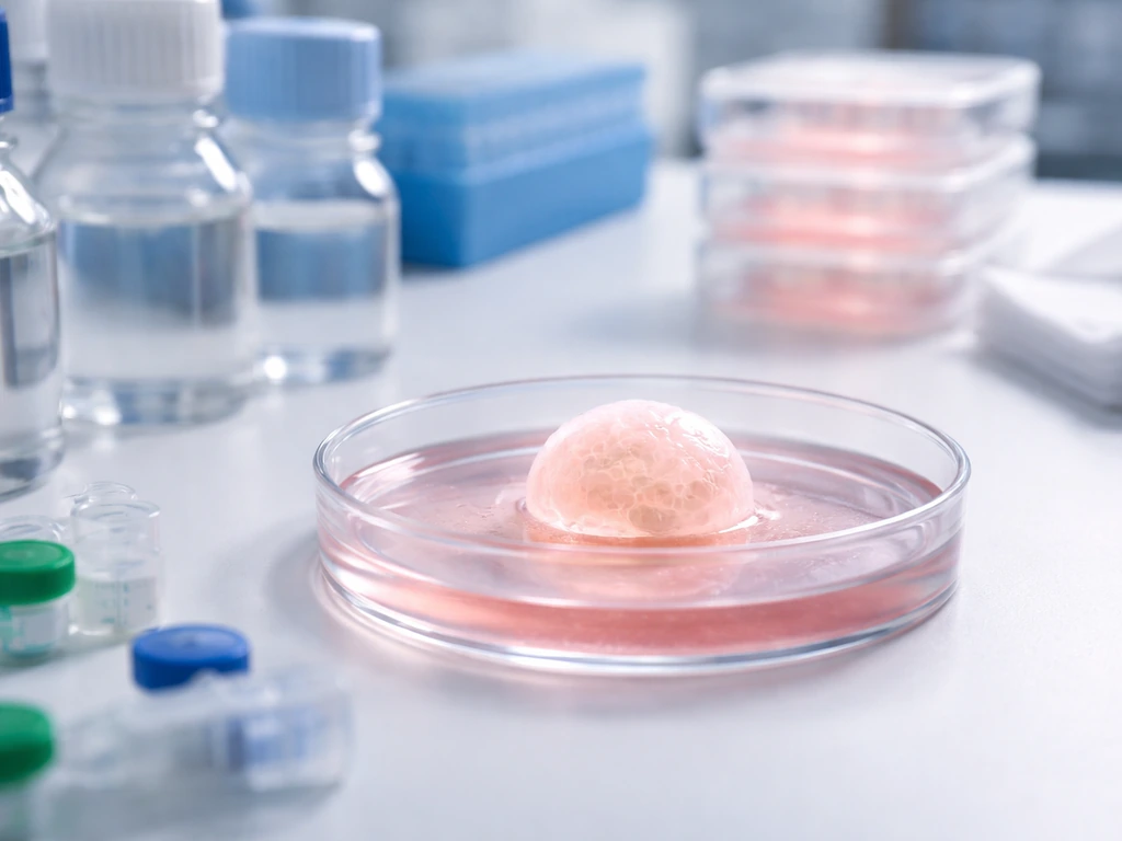
Yes, scientists can grow brain-like structures in a lab today, but not a complete, functional brain. What's actually possible right now is growing small 3D clusters of human brain cells called brain organoids, sometimes called mini-brains. These are typically 1 to 3 mm across, contain neurons that fire electrical signals and form synapses, and can survive in culture for many months. What's not possible is assembling a full, integrated brain with all its regions, wiring, blood supply, and higher-order function. That distinction matters a lot, because the two things are often confused.
Can You Grow a Brain in a Lab Today? What’s Feasible

Marcus Whitmore
2 Jun 2026
What people usually mean by 'growing a brain'
The phrase covers at least three different things, and they are not the same experiment. Understanding which one you mean changes the answer completely.
- Brain organoids: self-organizing 3D clusters of stem-cell-derived neurons and supporting cells that spontaneously develop structural features resembling early human brain development. These are the most common thing people are referring to when they say 'grow a brain in a lab.'
- 3D neuronal cultures: neurons grown in a gel or scaffold in three dimensions, often from iPSCs or primary cells, without necessarily self-organizing into distinct brain regions. More controlled, less complex than organoids.
- A whole functional brain: assembling a complete organ with all cortical layers, subcortical structures, a full vascular system, cranial nerves, and integrated network activity. This is not possible today and is not close.
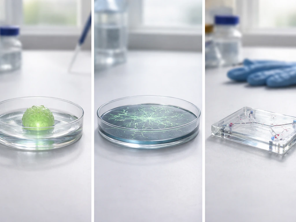
Brain organoids sit in an interesting middle ground. They are not just a random pile of neurons, but they are also nowhere near a real brain. Think of them as a rough, early draft of a small brain region, written in a dish, by cells that are trying to follow their own developmental instructions without most of the context they would normally have.
What we can actually do today
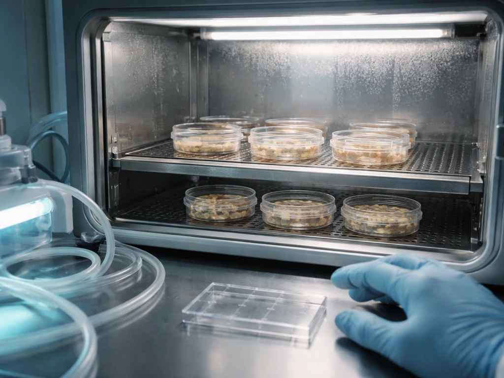
Current lab capabilities are genuinely impressive when you look at them clearly. Researchers can grow cerebral organoids that develop recognizable cortical-like layering, produce multiple neuron subtypes, and show spontaneous electrical activity. By around 3 months in culture, synaptic proteins like SYN1 appear, marking real synapse formation. By 8 months, some organoids generate spontaneously active neuronal networks and even include photosensitive cells that respond to light stimulation.
High-density electrode arrays can record coordinated electrical signals across 500-micron-thick organoid slices. One study using high-density CMOS microelectrode arrays reported spontaneous extracellular activity and mapped functional patterns across approximately 500 µm-thick human brain organoid slices [High-density electrode arrays can record coordinated electrical signals across 500-micron-thick organoid slices](https://pubmed. ncbi. nlm.
nih. gov/35906223/). Synchronized calcium transients, a sign of coordinated network communication, have been observed in human neural networks derived from organoids. That is real neuroscience happening in a dish.
But the gap between that and a brain is enormous. A real brain has roughly 86 billion neurons, a precisely organized vascular network delivering oxygen to every cubic millimeter, dozens of distinct regions with defined connectivity, and decades of development behind it. The largest organoids today top out around 3 to 4 mm in diameter. Beyond that, physics gets in the way, and we will get to that shortly.
It is also worth knowing that organotypic brain slice cultures exist as a separate experimental tool. These take actual slices of animal brain tissue and keep them alive in culture, preserving the 3D architecture of the original tissue. Useful for studying existing circuits, but fundamentally different from building a brain from scratch. Questions about what kinds of animal development should be allowed in artificial wombs also raise similar concerns about limits, safety, and ethical oversight.
How lab brain structures are actually made
Starting materials: where the cells come from
Most brain organoid work starts with induced pluripotent stem cells, or iPSCs. These are adult cells, often from skin or blood, that have been reprogrammed back to a stem-cell-like state. Because they can be made from any person, they carry that person's genetic identity, which makes them incredibly useful for studying disease. Embryonic stem cells can also be used. The key property is pluripotency: these cells can, in principle, become any cell type in the body, including neurons.
Differentiation: steering cells toward a brain identity
Getting stem cells to become brain cells requires guiding them through the same molecular decision points that happen during embryonic development. Researchers use specific combinations of growth factors, signaling molecules, and inhibitors added to the culture medium in a defined sequence. Understanding how researchers steer cell growth toward a brain identity helps explain how cells in our organs grow and develop specialized roles growth factors. The timing matters.
Cortical specification markers like TBR2 appear around day 21 in typical protocols. Generating a cerebral organoid with early brain-like organization takes roughly 10 days from initial seeding to basic structure, while obtaining cells with defined regional brain identity takes around 30 days. After that, organoids are maintained and monitored as they mature over weeks to months.
The 3D environment: scaffolds and matrix
Growing cells in two dimensions on a flat dish does not give you brain-like organization. For organoids, cells are typically embedded in or surrounded by an extracellular matrix material, most commonly Matrigel, which mimics the physical support tissue that surrounds cells in the body. This 3D environment allows cells to migrate, organize into layers, and develop spatial relationships that resemble actual brain tissue. Engineered hydrogel-based scaffolds are increasingly being explored as more defined, reproducible alternatives to Matrigel. The scaffold chemistry, stiffness, and composition all influence how cells behave, which is an active area of research.
The growth limits that stop organoids from scaling up
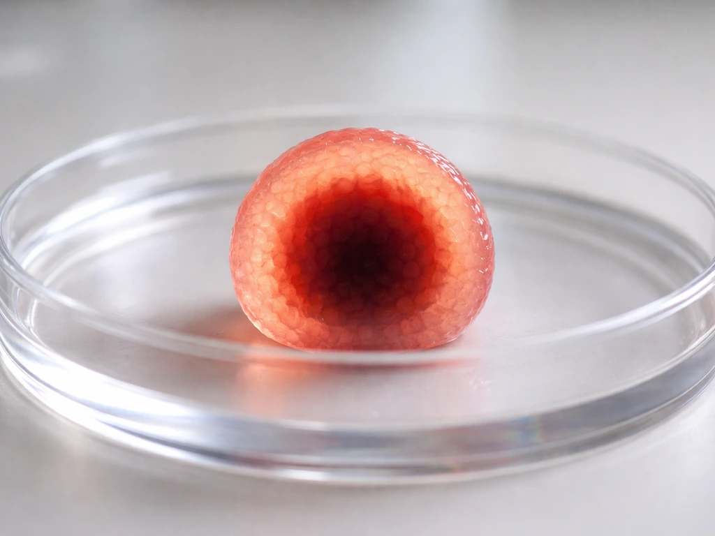
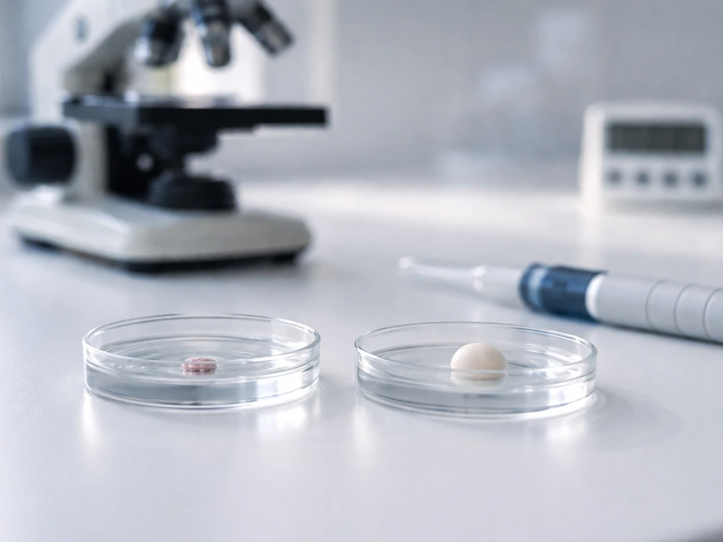
Here is where biology and physics join forces to stop you. The fundamental problem with growing anything bigger than a few millimeters without a blood supply is oxygen and nutrient delivery. In a living brain, every neuron is within about 100 micrometers of a capillary. In a dish, nutrients and oxygen can only reach cells by diffusing through the surrounding medium. That process becomes inefficient past roughly 200 to 400 micrometers from the surface. Organoids can grow to 3 or 4 mm in diameter, but the cells deep inside become oxygen-starved. By later culture time points, a necrotic, dead core is common. The outer shell is alive and active; the middle is not.
This is not a technical problem that better equipment will easily fix. It is a physical constraint rooted in how diffusion works, the same principle that limits how large a cell can grow before its surface area can no longer supply its volume. Solving it for a centimeter-scale or larger brain-like structure would require a functional vascular network, which is something that has not been achieved in lab-grown brain tissue. Researchers are experimenting with microfluidic systems and air-liquid interfaces to improve oxygen delivery to organoids, but these approaches address the outer layers, not true vascularization throughout the tissue.
Beyond oxygen, there are organizational constraints. A real brain is not just neurons packed together. It is a precisely wired structure where specific neurons in one region connect to specific neurons in another through long axonal projections. That wiring doesn't self-assemble spontaneously in a dish. The morphogen gradients, mechanical cues, and temporal signals that guide axons to their correct targets during real development are extraordinarily hard to replicate artificially.
How you measure whether it's actually working
Measuring success in organoid research means working through several layers of evidence, from basic cell survival up to coordinated network behavior. No single readout tells the full story.
| What you measure | How you measure it | What it tells you |
|---|---|---|
| Cell viability and identity | Immunostaining for neuron/glial markers, live/dead assays | Are the right cell types present and alive? |
| Structural organization | Histology, confocal imaging of layering and cell arrangement | Does the tissue resemble brain-region architecture? |
| Synaptic protein expression | Immunostaining for SYN1, PSD95, and similar markers | Are synapses forming? (SYN1 appears ~3 months) |
| Single-cell gene expression | Single-cell RNA sequencing, molecular mapping | What cell subtypes are present? How mature are they? |
| Calcium imaging | Fluorescent calcium indicators under a microscope | Are neurons active and communicating? (synchronized transients) |
| Electrophysiology | Multi-electrode arrays, CMOS microelectrode arrays, patch clamp | Do neurons fire action potentials? Are there network oscillations? |
| Longevity and reproducibility | Long-term culture, comparison across batches | How stable is the model? How consistent between experiments? |
One honest limitation to flag here: batch variability is a serious problem. Different organoid batches from the same starting cells can differ significantly in quality, size, and which brain regions they spontaneously develop. This 'batch syndrome' is well-documented and means that a single impressive result doesn't automatically mean reproducibility. Rigorous organoid science requires large numbers of replicates and careful characterization of each batch.
What is genuinely not possible yet
Growing a complete, functional brain in a lab is not possible today, and the barriers are not just technical gaps waiting for a better protocol. They are deep biological and physical challenges. There is no vascularization, so anything brain-sized would die at its core. There is no integrated connectivity between the dozens of distinct brain regions that define real brain function.
There is no immune system component, no meninges, no cerebrospinal fluid circulation. The complexity of development that produces a real brain involves not just the right cells, but the right spatial and temporal orchestration over years, in an environment (the skull, the body, with sensory input) that a lab dish cannot replicate. Similarly, transplanted organs do not simply grow larger by themselves; their growth depends on how well they integrate, receive oxygen, and remodel after surgery.
There is also a critical conceptual point: current brain organoids do not have consciousness. This reflects the prevailing scientific consensus backed by expert bodies including the ISSCR. Organoids lack the integrated complexity, connectivity, and sensory input that are thought to be prerequisites for conscious experience. The ethics literature is careful to note that this assessment should be revisited as organoids become more sophisticated, but right now, calling an organoid conscious is not supported by evidence. The worry is real enough that researchers and ethicists take it seriously, but the current state of the science does not warrant alarm on this front.
It is also worth noting that growing brain organoids is a fundamentally different question from growing other organs in the lab. That broader question, like can we grow organs, is different from making small brain-like structures in a dish. Questions about whether stem cells can grow organs more broadly, and whether scientists can grow organs for transplant, involve different constraints and timelines than what applies to brain tissue specifically. The brain's complexity, its demand for precise regional organization and long-range connectivity, makes it uniquely difficult even within organ engineering.
Practical next steps if you want to understand this field
Where to learn the actual science
The best starting point for serious learners is the primary literature, and a lot of it is freely accessible. The 2024 Nature Reviews Molecular Cell Biology paper on modeling human brain development with organoids is a strong overview of where the field sits. The ISSCR (International Society for Stem Cell Research) website is the go-to source for both the science and the governance framework. Their 2021 Guidelines for Stem Cell Research and Clinical Translation specifically cover organoids and provide a readable, authoritative framework. For the engineering side, Nature Reviews Bioengineering's 2023 piece on functional bioengineered CNS models gives a good picture of scaffold and design considerations.
What kind of lab setup enables this work
Brain organoid research requires a fully equipped cell biology and neuroscience lab. Key enabling technologies include iPSC culture infrastructure, biosafety cabinets, CO2 incubators, spinning bioreactors or orbital shakers to keep organoids in suspension, confocal microscopes for imaging, immunohistochemistry setups for staining, and ideally single-cell RNA sequencing access for molecular characterization. For functional readouts, multi-electrode arrays or high-density CMOS microelectrode arrays allow recording of electrical activity.
A PubMed review on electrophysiological analysis in brain organoids highlights electrophysiology, including multi-electrode arrays and probe-based recording, as a key frontier for functional characterization [multi-electrode arrays or high-density CMOS microelectrode arrays allow recording of electrical activity](https://pubmed. ncbi. nlm. nih.
gov/33510616/). Microfluidic platforms are increasingly used to address the oxygen delivery problem. This is not DIY territory: it requires trained personnel, institutional oversight, and significant infrastructure.
How to think about ethics and realistic expectations
If you are approaching this topic from an ethics angle, the key questions in the field right now center on moral status, consciousness assessment, and governance as organoids become more sophisticated. These same ethics questions also come up when people ask is it ethical to grow human organs in a lab. The ISSCR guidelines provide an ethics and oversight framework specifically designed for this research. Ethics papers in journals like the Journal of Medical Ethics and discussions comparing governance frameworks across countries (the US, UK, and Germany have all developed approaches) are worth reading if you want to engage seriously with this dimension.
The practical ethical guidance for researchers is: work within your institution's IRB and ethics board frameworks, use the ISSCR guidelines as a baseline, be careful with language (calling organoids 'mini-brains' invites public misunderstanding), and stay engaged with the evolving consensus on what advanced organoids may require in terms of oversight. For curious non-researchers, the main thing to calibrate is expectation: organoids are powerful, legitimate research tools that are teaching us enormous amounts about brain development and disease. They are not proto-brains, they are not conscious, and they are not a stepping stone to growing a replacement brain anytime soon.
The field is moving fast. What is impossible today may look different in five or ten years, particularly around vascularization and multi-region assembloid approaches where organoids representing different brain regions are fused together. If you want to stay current, following ISSCR updates and checking preprint servers like bioRxiv for organoid and neural engineering papers will keep you ahead of what makes it into textbooks.
FAQ
Can you grow a “brain” in a lab that can think or feel?
No. Even the most advanced brain organoids are not integrated into a whole-brain circuit, they do not have the sensory inputs, vascular support, and long-term developmental coordination that a living brain has, so there is no evidence they can experience perception or have consciousness.
How big can lab-grown brain organoids get, and why don’t they keep growing?
Most organoids are a few millimeters across (often cited around 1 to 3 mm, with top end around 3 to 4 mm). Past that scale, oxygen and nutrient diffusion limits typically produce a dead or non-viable core, so size growth hits a physical ceiling without true vascular function.
Do brain organoids have blood vessels, or can they be vascularized in the dish?
In general, organoids do not develop a functional, brain-wide vascular network that delivers oxygen throughout the tissue. Some engineering approaches may improve delivery near surfaces using microfluidics or air-liquid interfaces, but that is not the same as full vascularization throughout the organoid.
Are organoids just clumps of neurons, or do they form meaningful brain-like structure?
They are more structured than a random cell clump, because cells often form layered organization and multiple neuron subtypes, plus measurable electrical activity. Still, the organization is partial and not equivalent to the precise multi-region architecture and connectivity of a real brain.
What do researchers measure to show organoids are “working” versus just surviving?
Common indicators include synapse formation markers, coordinated electrical activity, and network-level readouts like calcium transients. Researchers also check for cell viability throughout the organoid, since deep-cell death can look like “failure” or can distort activity measurements.
Do brain organoids self-wire correctly over long distances like in a real brain?
Not reliably. Long-range axon targeting and precise inter-region connectivity are major unsolved challenges. The right timing, gradients, and mechanical cues that guide wiring during development are hard to reproduce in a dish.
Can I make a brain organoid at home or in a small lab?
No, it is not DIY territory. The process needs specialized incubator and sterile culture systems, appropriate expertise in handling stem cells, and tools for imaging and molecular characterization. Most importantly, lab safety and institutional oversight are required for human-derived cell work.
Why do organoids made from the same starting cells look different between batches?
This is a well-known “batch variability” problem. Even with the same protocol, batches can differ in size, maturity, and which brain region-like identities appear. That means conclusions should rely on multiple replicates and careful batch-level characterization.
Do organoids come from adult patients directly, and what does that affect?
Often they are derived from induced pluripotent stem cells (iPSCs) reprogrammed from adult cells, which preserves the donor’s genetic identity. That helps model disease, but it can also increase variability because different donors and reprogramming outcomes can change differentiation behavior.
What is the difference between brain organoids and brain slice cultures?
Brain slice cultures keep pieces of existing brain tissue alive in a controlled environment, preserving original architecture. Organoids instead build tissue from stem cells, so the developmental “construction” step and limitations are different.
If organoids show electrical activity, does that mean they can be “controlled” like a device?
Electrical signals alone do not imply controllability of complex function. While researchers can record activity using electrode arrays and sometimes stimulate responses, translating that into purposeful, integrated computation like a brain is far beyond current capabilities.
Does ethics concern mean organoid experiments are dangerous or unethical by default?
Ethical concern is not the same as saying the work is inherently unsafe or wrong. The field focuses on governance, appropriate oversight, and avoiding misleading public language. The consensus view is that current organoids lack consciousness, but oversight is meant to adapt if capabilities change.
Next Article
Is It Ethical to Grow Human Organs? A Practical Guide
Ethical, science-backed guide to growing organoids and engineered organs, covering consent, risk, justice, and limits.