Transplanted organs do not grow the way a child's kidneys grow during development. What actually happens is more nuanced: depending on the organ, a transplant can expand slightly through hypertrophy (individual cells getting larger), regenerate lost tissue (the liver is the standout here), or simply remodel itself to fit its new host without changing much in size at all. The short answer is that some transplanted organs do measurably increase in size or function after transplant, but this is driven by adaptation and repair, not the kind of programmed developmental growth that takes a newborn kidney to an adult one.
Do Transplanted Organs Grow? What Actually Happens After Transplant

Marcus Whitmore
11 Apr 2026
Do transplanted organs actually grow in size?
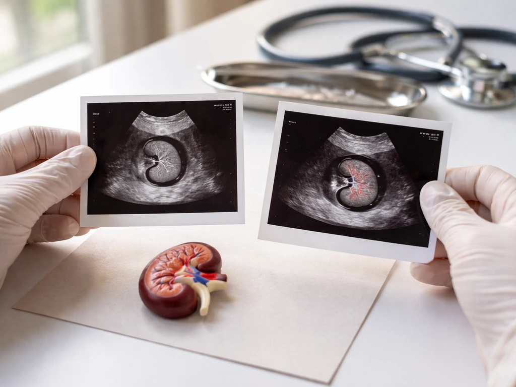
Yes, but with important caveats. The clearest example is the kidney. Research tracking renal allograft length by ultrasound at transplant and then at 1, 3, 6, and 12 months found that 57.7% of recipients showed a mean increase in kidney length of about 0.3 cm within the first month after transplant. That is compensatory renal hypertrophy: the transplanted kidney senses it is now the sole functioning kidney in the body and responds by enlarging its existing cells and ramping up filtration capacity. It is not growing new nephrons; it is making the ones it has work harder and, to a modest degree, physically bigger.
The liver behaves differently and more dramatically. In living-donor liver transplants, where only a partial liver is transplanted, real regenerative growth happens quickly. Graft volume as a proportion of standard liver volume increases rapidly over the first few months after transplant. The liver is the body's most regenerative solid organ, and that capacity does not disappear just because the tissue has been moved into a new person. This is about as close as transplant medicine gets to genuine post-transplant growth.
The heart and lungs are a different story. These organs have very limited regenerative capacity. After a heart or lung transplant, you are not going to see meaningful size increases driven by healthy adaptation. What you see instead is remodeling, and not always the good kind.
Why some transplanted tissues adapt (and what that actually looks like)
The reason some transplanted organs can adapt at all comes down to how cells in our organs grow and respond to physiological signals. In the kidney, the process is driven by glomerular hyperfiltration: when one kidney is removed from a donor and placed into a recipient who had two failing kidneys, the transplanted organ suddenly faces a higher filtration demand than it was designed for as one of a pair. Single-nephron adaptations, including increased blood flow and higher filtration pressure per glomerulus, push the organ toward compensatory hypertrophy. Cells in the tubules and glomeruli get larger, and the organ measurably increases in size over months.
In the liver, adaptation is not just hypertrophy. It involves actual cell proliferation, where hepatocytes divide to restore lost mass. Research on living-donor liver transplantation shows that this regenerative ability can be maintained even when early allograft dysfunction occurs, as long as the graft-to-recipient weight ratio (GRWR) is adequate. Think of it like a sourdough starter: as long as the base culture has enough viable material and the right environment, it will double and expand. Cut it too small and recovery stalls.
Cell turnover also plays a role across all transplanted organs, just at slower rates. Over months and years, the donor cells in a transplanted organ are gradually replaced by new cells generated through normal tissue maintenance. In some organs, this turnover is extensive enough that the organ becomes partly composed of cells that were never part of the original donor tissue, though the structural architecture stays the same.
Why the body puts a ceiling on post-transplant growth
Even in organs capable of adaptation, growth is tightly constrained. The most immediate limit is vascular supply. A transplanted kidney, for example, is connected to a fixed set of blood vessels in the recipient's pelvis. Its growth is physically bounded by how much oxygen and nutrients those vessels can deliver. Research on renal blood flow shows that as kidney function declines, renal blood flow drops substantially, from around 1.24 ml/min/g in healthy-function kidneys down to roughly 0.53 ml/min/g in severely impaired ones. No perfusion, no growth.
Beyond blood supply, the body uses homeostatic signaling to sense when organ size and function are sufficient. Once a transplanted kidney achieves an adequate filtration rate for the recipient's body size, the hypertrophic stimulus diminishes. The organ does not keep expanding indefinitely, any more than a muscle keeps growing after you stop adding training load. Mechanical constraints from surrounding tissue and the organ's own structural architecture also set physical limits on how much cell swelling can occur before function is compromised.
For the liver, inadequate graft mass is a real clinical problem. Small-for-size syndrome, where the transplanted liver remnant is too small relative to the recipient's metabolic needs, can drive graft failure. The organ tries to regenerate but cannot keep up with demand, and the result can be catastrophic dysfunction requiring retransplantation. This illustrates perfectly why growth after transplant is not unlimited: the biological signals say "grow," but the physiological ceiling says "only so fast, only so much."
How different organs behave after transplant
Not all transplanted organs have the same capacity for adaptation or size change. Here is a breakdown of the major ones:
| Organ | Adaptive capacity | Main mechanism | Typical size change post-transplant |
|---|---|---|---|
| Kidney | Moderate to strong | Compensatory hypertrophy, glomerular hyperfiltration | Measurable increase (~0.3 cm length) within first months |
| Liver (partial) | Very strong | True cell proliferation and regeneration | Rapid volume increase over first 1–6 months |
| Heart | Limited | Reverse remodeling (shrinkage), not enlargement | Left ventricular mass typically decreases after kidney transplant |
| Lungs | Very limited | Structural remodeling (often fibrotic) | No healthy enlargement; fibrotic restriction is a major concern |
| Liver (whole) | Moderate | Hypertrophy and cell turnover | Modest adaptation to recipient metabolic demands |
The heart deserves a special note here. When researchers tracked echocardiographic changes after kidney transplantation, they found that left ventricular mass index actually decreased between 1 month and 12 months, with further decline by 24 months.
In the review evidence on reverse remodeling after kidney transplantation, echocardiographic studies commonly report regression of left ventricular mass index, including a prospective cohort (143 recipients) in which left ventricular mass index fell between 1 and 12 months and declined further by 24 months [left ventricular mass index actually decreased between 1 month and 12 months, with further decline by 24 months](https://pmc. ncbi. nlm. nih.
gov/articles/PMC12958384/). An echocardiographic study in kidney transplant recipients assessed cardiac structural and functional recovery, including changes such as left ventricular hypertrophy and left ventricular ejection fraction around 6 to 12 months after transplantation [left ventricular mass index actually decreased between 1 month and 12 months](https://pubmed. ncbi. nlm.
nih. gov/29416201/). The heart was not growing, it was recovering from the strain of chronic kidney disease. This is a good example of reverse remodeling: the transplanted kidney reduces the hemodynamic burden on the heart, and the heart shrinks toward a healthier size.
The transplanted organ growing and the recipient's own organs remodeling are two separate phenomena that can happen simultaneously.
What happens over time: early adaptation vs. long-term changes
The first weeks and months
Early post-transplant remodeling is mostly about the organ establishing function in its new environment. For kidneys, the hypertrophic response begins almost immediately, with measurable size increases within the first month. For partial liver grafts, regeneration kicks in rapidly as the liver responds to the metabolic demands of the recipient's body. During this window, the organ is also dealing with surgical stress, ischemia-reperfusion injury (the damage from having blood flow cut off during harvesting and restored after transplant), and the early phases of the recipient's immune response.
Months to years: stabilization and slower drift
After the initial adaptation phase, changes become slower but do not stop. Kidney volume and function measurements show continued change at follow-up times of 6 months, then again years later, with documented changes at median follow-up periods extending to around 6 years. The organ is in a slow, ongoing dialogue with the host's physiology. Long-term, this can go two ways: stable functional adaptation, or progressive decline driven by cumulative injury and fibrosis.
For the liver, long-term changes often involve idiopathic post-transplant hepatitis or chronic antibody-mediated rejection, both of which can cause progressive fibrosis that gradually replaces healthy regenerated tissue with scar. Whether we can grow organs to avoid these chronic injury patterns is one of the driving questions in regenerative medicine today.
When growth goes wrong: rejection, scarring, and chronic injury
Not all growth-like changes in a transplanted organ are beneficial. The immune system's response to the foreign graft can drive harmful remodeling that mimics some aspects of growth but is actually destructive. TGF-beta signaling in T cells plays a central role here: it is required for the fibrosis associated with chronic rejection, linking immune activity directly to the scar tissue formation that strangles graft function over time. Elevated TGF-beta and PDGF-B, along with increased collagen deposition, are hallmarks of chronic allograft nephropathy, particularly when hypertension is present. This is profibrotic remodeling, not healthy enlargement.
In kidney transplant biopsies, inflammation in areas of fibrosis (called i-IFTA) is strongly associated with decreased long-term graft survival. The Banff classification system, which pathologists use to grade transplant rejection on biopsy, flags i-IFTA alongside other histological criteria as a marker of chronic active T-cell-mediated rejection. These findings confirm that scar-driven remodeling is not a growth process in any useful sense. It is the opposite: it is progressive loss of functional tissue replaced by non-functional connective tissue.
Chronic lung allograft dysfunction (CLAD) is the lung-transplant equivalent. It comes in two main forms: bronchiolitis obliterans syndrome (BOS), which is obstructive and progressive, and restrictive CLAD, which involves fibrotic changes to the airway and parenchyma. Research shows that bilateral lung recipients with CLAD experience the greatest losses in FEV1 and FVC during the first 6 months after CLAD onset. These are measurable functional declines tied directly to fibrotic remodeling, and they are largely irreversible.
Vascular occlusion adds another layer of complexity. When rejection-driven inflammation narrows graft blood vessels, ischemia follows, and ischemic tissue undergoes necrosis rather than repair. The resulting fibrosis further restricts the growth signals that might otherwise support healthy adaptation. It becomes a self-reinforcing cycle of damage. This is the biological reason that questions about whether it is ethical to grow human organs for transplantation carry real urgency: better-matched or lab-grown organs could reduce the immune conflict that drives these destructive changes.
How doctors measure and monitor changes after transplant
The practical question for anyone going through or studying a transplant is: how do clinicians actually track whether an organ is growing, remodeling, or failing? The answer depends on the organ, but there are consistent principles across all transplant types.
Imaging
Ultrasound is the workhorse for kidney transplant monitoring. Serial ultrasound measurements of kidney length and volume over 6 to 12 months can track hypertrophic changes, and Doppler ultrasound parameters measured in the first week after transplant, including the resistive index, predict graft function at 6 months. Contrast-enhanced ultrasound (CEUS) adds information about perfusion: patients with impaired graft function show different resistive index values and perfusion patterns from those with preserved function. For liver transplants, grayscale and color Doppler ultrasound are the standard early post-op tools, with contrast-enhanced CT used to evaluate complications like hepatic artery thrombosis.
Blood markers and functional tests
For kidneys, estimated GFR (eGFR) is the primary functional measure. It reflects how well the organ is filtering, which correlates with both healthy hypertrophy and harmful fibrosis. For livers, serum bilirubin, liver enzymes, and synthetic function markers (like INR) track both regeneration success and rejection. For lungs, spirometry is the cornerstone. FEV1 decline criteria are used to detect both chronic rejection (BOS is defined partly by a sustained FEV1 drop to below 80% of best post-transplant value) and acute respiratory events in home monitoring programs. Some centers now use home spirometry with defined FEV1 decline thresholds to catch early deterioration before it becomes irreversible.
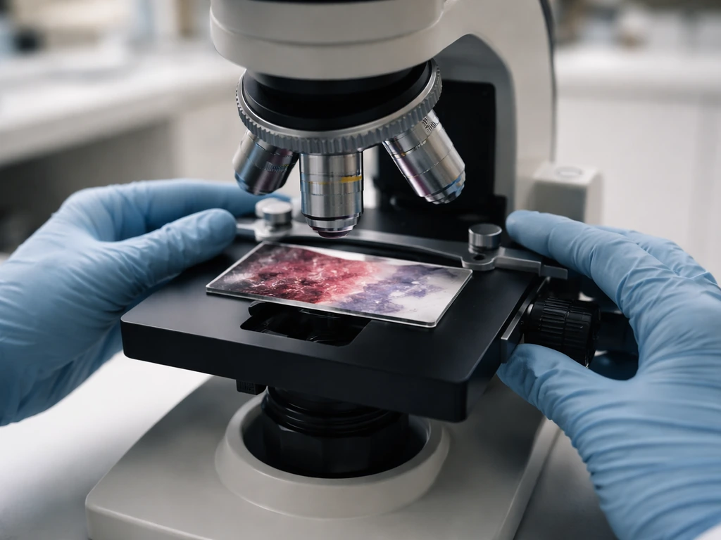
Biopsy
When imaging and blood markers are not enough, biopsy provides ground truth. In kidney transplants, protocol biopsies and indication biopsies assess fibrosis burden, inflammation patterns (including i-IFTA), and signs of rejection using the Banff classification. In liver transplants, serial surveillance biopsies have been used in some centers to track long-term fibrosis development from sources like recurrent viral hepatitis or chronic antibody-mediated rejection. Biopsy is invasive, so it is generally reserved for cases where other tests leave the diagnosis uncertain or treatment decisions hinge on knowing the exact histological picture.
The bigger biology picture: growth, repair, and constraints
Zooming out, what happens in transplanted organs is a compressed version of the same growth biology that operates across all living systems. Growth in biology is never unlimited; it is always the result of a signal, a cellular response, and a constraint that eventually wins. Whether stem cells can grow organs in ways that avoid these constraints is an active area of research. Scientists working on growing organs in the lab are trying to engineer tissues that have better regenerative potential and reduced immunogenicity, precisely because they understand how current transplanted organs hit their physiological ceiling.
The transplanted kidney enlarging slightly because it is the only kidney is the same logic as a plant root growing toward water: the system detects a deficit, mobilizes a growth response, and scales up until the environment says stop. The difference is that in transplant medicine, "the environment saying stop" can mean fibrosis, rejection, or inadequate blood supply, rather than a soft soil boundary. Understanding those constraints is exactly why researchers study how scientists grow enough cells for their research into transplant biology, and why pushing past those constraints is one of the central challenges in the field.
There are also broader questions on the horizon. Whether scientists should be allowed to grow animals in artificial wombs as a source of transplantable organs touches on the same biological principles: can we create an organ with the right growth trajectory, the right architecture, and the right cellular machinery to survive and adapt in a human body? And at the far edge of the question, whether you can grow a brain in a lab illustrates just how far the science of organ growth still has to go before transplant medicine can fully harness it.
What this means practically
If you are a student trying to understand transplant biology, here is the takeaway: transplanted organs change in size and function after transplant, but the direction and mechanism depend almost entirely on the organ type, the match between donor and recipient, the immune environment, and the adequacy of blood supply. Kidneys hypertrophy modestly. Partial livers regenerate substantially. Hearts and lungs mostly remodel, and not always for the better. The body does not simply "grow" a transplanted organ to fit; it negotiates between the organ's own biology, the recipient's physiology, and the ongoing immune standoff that defines the post-transplant life of every graft.
- Expect kidney transplants to increase slightly in size within the first month due to compensatory hypertrophy, measurable by ultrasound.
- Partial liver grafts genuinely regenerate over 1 to 6 months, but graft mass relative to recipient size must be adequate or failure can result.
- Heart and lung transplants do not grow; adverse remodeling (fibrosis, BOS) is the main concern over time.
- Long-term fibrosis driven by chronic rejection, hypertension, or recurrent injury is the primary threat to all transplanted organs.
- Monitoring combines imaging (ultrasound, CT), functional tests (eGFR, spirometry, liver enzymes), and biopsy when needed.
- Growth after transplant is always constrained by vascular supply, immune response, and homeostatic signaling.
FAQ
Do transplanted kidneys grow new filtering units after surgery?
They do not typically create new nephrons. The main early change is compensatory hypertrophy, where existing nephrons enlarge and increase filtration. Over the long term, function can still improve or decline depending on whether chronic injury is controlled, but the tissue is not “rebuilding from scratch.”
If an organ increases in size, does that always mean the transplant is doing well?
Not necessarily. Size gain can reflect healthy adaptation, but similar outward changes can also accompany harmful processes like inflammation and fibrosis, especially when immune injury is ongoing. Clinicians usually interpret growth alongside functional markers (kidney eGFR, liver synthetic function, and lung spirometry) and, when needed, biopsy findings.
How long does “growth-like” adaptation last after a transplant?
For kidneys it is most evident in the first months, with measurable changes continuing later (including years). For partial livers, regeneration is rapid after transplant but long-term outcomes depend on complications like rejection and recurrent hepatitis. Hearts and lungs generally show slower remodeling rather than true enlargement driven by healthy regeneration.
Can donor organ size affect whether the transplant can “grow” enough?
Yes. In liver transplants, especially, the graft-to-recipient weight ratio matters, because being too small can trigger small-for-size syndrome. When the graft cannot meet metabolic demand, regeneration may not catch up, leading to dysfunction rather than beneficial growth.
Does better blood flow always lead to more adaptation?
Adequate perfusion is necessary, but it is not the only requirement. Even with good early blood supply, ongoing immune injury can push the organ toward scarring instead of functional expansion. Also, if perfusion drops, the organ can lose the biological conditions needed to sustain healthy hypertrophy.
What causes the heart to get smaller or stop “growing” after transplant?
In many kidney-to-heart scenarios, the heart’s left ventricular size decreases because the kidney transplant improves overall fluid and blood pressure balance. This is reverse remodeling of the recipient’s heart, not growth of the heart graft, and it depends on stabilizing the underlying chronic disease that strained the heart.
Why don’t lung transplants show meaningful size increase over time?
Because lungs have limited regenerative capacity once significant structural injury or airway remodeling occurs. After transplant, chronic lung allograft dysfunction tends to produce progressive fibrotic changes, and functional decline (such as sustained FEV1 loss patterns) reflects scarring more than beneficial enlargement.
Is it possible for transplanted organs to be partly made of the recipient’s cells?
Yes, in some organs there can be substantial cell turnover over months to years, meaning portions of the tissue may include cells that were generated after transplant rather than only the original donor cells. The organ’s architecture may remain similar even while the cellular composition shifts.
How do clinicians distinguish healthy hypertrophy from harmful chronic rejection?
They combine trends in function tests with imaging and, when needed, biopsy. For kidneys, fibrosis and inflammation patterns tied to chronic rejection (including metrics like i-IFTA) help separate “doing more work” from “losing functioning tissue.” For livers, enzyme and synthetic function trends plus targeted evaluation for rejection or recurrent disease guide interpretation.
If my transplant seems to be growing, when should I be concerned about complications?
Concern is warranted if growth is paired with rising inflammatory signals, worsening function tests, new vascular issues, or imaging patterns that suggest reduced perfusion. A key red flag is functional deterioration despite apparent size changes, because that pattern suggests remodeling or scar formation rather than true recovery.
Next Article
Can Stem Cells Grow Organs? What’s Possible and How
Yes for organoids and tissue-like structures from stem cells; full, vascularized, wired organs are still largely unreal.