Scientists grow enough cells for research by combining the right cell source, optimized culture conditions, and a staged expansion plan that works with the biology of cell division rather than against it. In practice, that means starting with a small, healthy seed population, splitting cultures before they get too crowded, scaling through progressively larger vessels, and tracking viability and cell number at every stage until you hit your target quantity with the function intact.
How Do Scientists Grow Enough Cells for Research?

Marcus Whitmore
5 May 2026
What 'enough cells' actually means before you start
Before you touch a flask, you need a concrete number in mind. 'Enough' has three dimensions: count, viability, and function. Count is the raw number your assay requires (a drug screen might need 10,000 cells per well across 96 wells, which is already 1 million cells before you account for dead cells or pipetting losses). Viability is the percentage of those cells that are actually alive, typically measured by trypan blue exclusion or a fluorescent viability assay. Most protocols require at least 85–90% viability at the point of use. Function is the hardest to pin down but the most important: do your cells still express the receptor, enzyme, or behavior you're studying? A million dead or dedifferentiated cells is scientifically worthless.
A useful rule of thumb is to calculate your minimum experimental requirement, then multiply by at least 1.5 to 2 as a buffer for losses during harvest, counting error, and any failed wells. Write that target number on the whiteboard before you seed the first plate. It becomes the anchor for every decision that follows.
The cell biology that limits (and enables) growth
Cells don't just divide on command. Every mammalian cell runs through a tightly regulated cell cycle: G1 (growth and preparation), S phase (DNA replication), G2 (checking that replication worked), and M phase (mitosis, where the cell physically splits in two). Checkpoints at each transition prevent a damaged or under-resourced cell from dividing. That's actually good news in the lab because cells in good conditions clear those checkpoints efficiently and divide at a predictable rate.
The main biological brakes on expansion are worth knowing because you'll hit all of them eventually. Contact inhibition stops many normal cell types from dividing once they physically touch neighbors, which is why a confluent monolayer essentially stops growing. Nutrient and waste gradients build up as density increases: glucose depletes, lactate and ammonia accumulate, and pH drops. Cells also respond to spatial constraints, not just chemical ones. Think of it like a garden bed: you can keep planting seeds, but once the bed is full, nothing new can get established without pulling something out first.
There's also the longer-term limit of replicative senescence. Normal (non-immortalized) primary cells have a finite number of divisions they'll complete before permanently exiting the cell cycle, roughly described by the Hayflick limit. This isn't a problem for the first few passages, but it becomes critical for multi-week expansion projects. Immortalized cell lines bypass senescence through mutations in cell cycle checkpoints, which is why they're workhorse lines in many labs, though that same property means you need to watch for genetic and phenotypic drift over time.
Choosing your cell source and culture format
Primary cells vs. immortalized lines
Primary cells are freshly isolated from tissue. They behave most like cells in the body, which makes them ideal for physiology-focused research. The trade-off is that they're finite: they arrive in small numbers, have limited passage capacity, and can be expensive and variable between donors. Immortalized cell lines (HeLa, HEK293, CHO, and dozens of others) divide indefinitely, grow fast, and are highly reproducible batch to batch. If your experiment requires enormous cell numbers or frequent repetition, an immortalized line is usually the practical choice.
Adherent vs. suspension culture
Adherent cultures grow attached to a surface, typically plastic treated for tissue culture. Most primary cells and many immortalized epithelial or fibroblast lines are naturally adherent. Suspension cultures grow in liquid without attachment and are typical for blood-lineage cells and some immortalized lines like Jurkat or CHO adapted for suspension. The format has huge practical consequences for scaling: adherent cultures are limited by surface area (more cells = more flasks or larger multi-layer vessels), while suspension cultures scale more easily in stirred bioreactors or spinner flasks.
| Feature | Adherent Culture | Suspension Culture |
|---|---|---|
| Cell types | Epithelial, fibroblast, neurons, most primary cells | Hematopoietic cells, some adapted lines (CHO, Jurkat) |
| Scale-up method | More surface area (larger flasks, multilayer vessels) | Larger volume (spinners, bioreactors) |
| Harvesting | Requires detachment (trypsin/EDTA) | Centrifugation, no detachment needed |
| Physiological relevance | High for tissue-derived cells | High for blood/immune cells |
| Scalability | Moderate; becomes labor-intensive at large scale | High; volume-driven, easier to automate |
The conditions that actually drive cells to expand
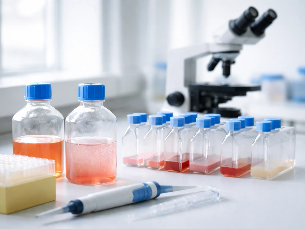
Culture medium is the foundation. Most mammalian cells use a base medium like DMEM, RPMI-1640, or MEM, which supplies glucose, amino acids, vitamins, and buffering capacity. To that base you add serum (usually fetal bovine serum at 5–20%) for growth factors, hormones, and attachment proteins, unless you're using a defined serum-free formulation. Many specialized cell types also need specific supplements: L-glutamine as an energy and biosynthesis source (it degrades spontaneously, so it's often added fresh), non-essential amino acids, antibiotics if contamination risk is high, and cell-type-specific growth factors like EGF, FGF, or insulin.
Gas environment matters as much as media. Mammalian cells are cultured at 37°C in a humidified incubator with 5% CO2, which keeps the bicarbonate-based buffer system in the medium at physiological pH (around 7. For bicarbonate-buffered culture media in a standard 5% CO2 incubator, bicarbonate-buffered media exhibit pH around 7.4 standard 5% CO2 incubator keeps bicarbonate-buffered media near physiological pH (around 7.4). 2–7.4). Oxygen is typically atmospheric (~21%) for most lines, though some specialized cultures (stem cells, tumor hypoxia models) use lower O2 levels. Letting a culture sit open on the bench for too long, or forgetting to replace the incubator's water pan, will stress your cells in ways that aren't immediately obvious but show up later as slower doubling times and increased spontaneous death.
Surface and coating choices matter for adherent cells. Standard tissue-culture-treated plastic works for most lines. More demanding cell types (neurons, primary hepatocytes, some stem cells) need extracellular matrix coatings like fibronectin, laminin, collagen, or Matrigel to attach properly and maintain function. Getting the coating wrong is a common reason primary cells fail to expand despite otherwise good conditions.
Scaling up step by step
Expansion is a staged process. You start small, confirm the cells are healthy and growing, then incrementally move to larger vessels. Trying to jump straight from a vial of frozen cells to a ten-layer flask is a reliable way to lose the entire stock.
- Thaw and recover: Revive your frozen stock into a T-25 flask with fresh, pre-warmed medium. Let cells settle and attach overnight before the first medium change. At this stage, your only goal is viability, not expansion.
- Establish a healthy culture: Once the T-25 is 70–80% confluent and cells look morphologically normal, passage into a T-75. This is where you confirm your doubling time and make sure the cells are actually in log-phase growth.
- First expansion stage: From the T-75, split into two or three T-75s or one T-175. Keep 80–90% confluence as your subculture trigger. Splitting too late (at 100% confluence) pushes cells into contact inhibition and degrades growth in subsequent passages.
- Second expansion stage: From healthy T-175s, move into multi-layer flasks (e.g., CellSTACK, HYPERFlask), roller bottles, or spinner flasks if your cells tolerate it. Each vessel type multiplies the effective surface area or volume without multiplying your labor proportionally.
- Final harvest: Time the last passage so cells reach your target density close to your experiment date. Harvest by trypsinization (adherent) or centrifugation (suspension), count with a hemocytometer or automated counter, confirm viability, and proceed to your assay.
For very large-scale needs, stirred-tank bioreactors or hollow-fiber bioreactors become the practical tools. These are more common in bioprocessing (vaccine or biologics production) but are increasingly accessible in academic settings for projects that genuinely need tens of billions of cells, as sometimes required for cell therapy work. Understanding how cells grow in organs and how transplanted or lab-grown tissue behaves at scale connects directly to this engineering challenge. That same question also raises important ethical concerns when discussing whether it is ethical to grow human organs how transplanted or lab-grown tissue behaves. Do transplanted organs grow because the graft can integrate and proliferate in its new environment, or do they mostly function without expanding much?
Planning and doing the math
The single most useful number you can know about your cell line is its doubling time: how many hours it takes for the population to double in log-phase growth. HEK293 cells double roughly every 20–24 hours. Primary human fibroblasts may take 36–48 hours. Some primary cells double even more slowly. You can calculate doubling time empirically by seeding a known number of cells, counting at regular intervals for two to three days, and fitting the data to an exponential growth curve.
With doubling time in hand, you can work backwards from your target. If you need 100 million cells in 10 days, and your line doubles every 24 hours, you theoretically need to start with about 100,000 cells (roughly 10 doublings would yield ~100 million). In reality, you add overhead for lag phase after thawing, suboptimal seeding densities, and passaging losses, so experienced researchers often start with 3 to 5 times the theoretical minimum.
Seeding density is just as important as total cell number. Most adherent lines have an optimal seeding density, often listed in the supplier's protocol, that gives cells enough neighbors to feel 'settled' but enough space to proliferate. Seeding too sparse causes slow recovery and lag; seeding too dense causes premature confluence and poor expansion. A common starting point for many adherent lines is around 2,000–10,000 cells per cm2, scaled to your flask surface area.
Keep a passage log. Write down the date, flask size, cell count at harvest, viability, and passage number every single time. This takes two minutes and saves hours of troubleshooting when something goes wrong three weeks later.
Quality control and fixing things when they go wrong
Contamination
Bacterial or fungal contamination is visible: your medium turns cloudy or yellow, and you'll often see a haze under the microscope. Discard the culture immediately and do not try to rescue it with antibiotics. Mycoplasma contamination is invisible to the eye and microscope, but it silently alters proliferation, gene expression, and cell metabolism. In lab troubleshooting discussions, people also flag that PCR readouts can be tricky and discuss controls and interpretation when screening for mycoplasma PCR false positives blank" rel="noopener noreferrer">mycoplasma contamination is invisible to the eye and microscope, but it silently alters proliferation, gene expression, and cell metabolism. Test for mycoplasma at least every three months on any line you're actively using, especially before a major experiment. Commercial PCR-based test kits take a few hours and are cheap insurance.
Viability decline
If viability is dropping across passages, the first things to check are medium age (has the glucose or glutamine been depleted?), pH (is the medium turning yellow before you change it?), and thawing protocol (was the cryoprotectant like DMSO removed quickly enough after thaw?). Cells that were never quite happy after thawing will compound the problem with each passage. When in doubt, go back to frozen stock early rather than trying to nurse a struggling culture back to health.
Senescence and phenotypic drift
Primary cells approach senescence after a finite number of passages, typically showing enlarged, flattened morphology, slow growth, and positive staining for senescence-associated beta-galactosidase. Keep passage numbers as low as possible by using sufficient starting material. Immortalized lines don't senesce but can drift genetically, especially if grown under selective pressure or at high passage numbers. Maintain a low-passage working bank in liquid nitrogen and refresh regularly. If your cells start behaving differently than your historical controls, check the passage number first.
Phenotypic and functional checks
At key expansion milestones, confirm your cells are still what you think they are. That might mean a morphology check under the microscope (are they still the right shape and size?), flow cytometry for surface markers, or a quick functional assay. This is especially critical if you're working with stem cells or primary cells where identity is closely tied to culture conditions. The question of how cells in our organs grow, and how lab-grown populations mirror or diverge from that behavior, is one of the more fascinating bridges between basic biology and applied cell culture. Those same principles are also central to the question can we grow organs from cultured cells in a way that behaves like real tissue how lab-grown populations mirror or diverge from that behavior.
When to rethink your strategy entirely
Sometimes the honest answer is that your current approach won't get you to your target. If you're hitting biological limits (primary cells senescencing, slow doubling times making timelines impractical) or logistical limits (no space for 50 T-175 flasks), it's time to consider alternatives. Three options come up most often:
- Switch cell type: If primary cells are too limiting, consider whether a validated immortalized surrogate would answer the same scientific question. Many labs run initial experiments in an immortalized line, then confirm key findings in primary cells.
- Optimize before scaling: If your cells are growing slowly or dying, fixing the underlying condition problem (media formulation, coating, CO2 balance) will pay off far more than just adding more flasks.
- Outsource or collaborate: Contract research organizations (CROs) and core facilities can expand cells at scales most academic labs can't match, often at lower total cost than the researcher time involved in doing it in-house. This makes most sense for single large-scale runs rather than ongoing needs.
- Switch to a bioreactor or 3D system: For very large cell numbers or for projects where 3D organization matters (organoids, spheroids, tissue-engineering work), the flat-flask paradigm may simply be the wrong tool. Microcarrier-based bioreactors can expand adherent cells in suspension, dramatically increasing yield per unit of lab space.
The broader question of whether and how scientists can grow organized tissues and organs from cultured cells, touching on topics like stem cell-derived organ growth, connects directly to these scale-up challenges. This is why stem cell biology is often paired with organ-level engineering goals stem cell-derived organ growth. Growing cells in large numbers for a basic biochemistry assay and growing cells to form a functional tissue are on the same biological continuum, just with very different engineering requirements. While growing cells is a core first step, creating a brain-like structure in a lab is a much bigger, specialized challenge that depends on cell types, scaffolds, and development-like signaling lab-grown populations. Researchers also ask whether scientists should be allowed to grow animals in artificial wombs, given the ethical and safety implications.
Your practical starting checklist
If you're setting up a cell expansion run right now, work through these steps before you open the incubator door:
- Define your target: final cell number, minimum acceptable viability (85–90% is a common standard), and the functional marker you'll check at harvest.
- Know your doubling time: look it up from literature, your supplier's datasheet, or measure it yourself in the first passage after thaw.
- Calculate your expansion plan: work backwards from the target number, account for seeding density requirements, passaging losses (~20–30% per split is a reasonable estimate), and your available timeline.
- Choose your vessel format: match the scale to the cell type (adherent vs. suspension) and your lab's equipment.
- Set your subculture trigger: for adherent monolayers, subculture at 80–90% confluence to keep cells in active log-phase growth and prevent contact inhibition from stalling expansion.
- Log everything: date, passage number, cell count, viability, medium used, any anomalies.
- Test for mycoplasma before your final experiment if you haven't done so in the past three months.
- Have backup frozen stock ready: if a culture fails at passage 5, you want to restart from passage 2, not from zero.
Cell expansion looks straightforward until the first time a culture crashes two days before your experiment. The researchers who reliably produce clean, high-viability cell preparations aren't doing anything magical. They're planning carefully, tracking obsessively, and respecting the biology instead of fighting it.
FAQ
How do scientists define “enough cells” when the assay depends on cell state, not just cell number?
Use your required cell count to back-calculate “start cells,” but also define what “function” means in your experiment (for example, percentage positive for a marker, enzymatic activity, or uptake/response). If function is measured by a readout that depends on activation state, schedule passaging and harvest timing so cells spend enough time in the correct growth phase before the assay.
What should I do if viability drops as I scale up, even though the early passages look healthy?
Don’t rely on a single viability check early in expansion. Re-measure viability and cell phenotype at every passage milestone, especially right before the final scale-up step. If a line keeps losing viability late, the issue is often not the initial inoculation but accumulation of waste or suboptimal passaging timing.
How can I troubleshoot cultures that hit confluence earlier than the doubling-time calculations predict?
If cells are reaching confluence sooner than expected, lower the seeding density target for the next run or shorten the time between passage/harvest to keep cultures out of the contact-inhibited, nutrient-depleted zone. Also verify you are using the correct growth surface area (flask type, multilayer vessel specs), since mis-scaling surface area is a common source of “too fast to confluence” problems.
Why do my cell counts look inconsistent, and how do I reduce counting error during expansion?
Confirm you are counting the right thing. Clumps inflate the measured count, while debris inflates dead-cell estimates. Use a consistent dissociation approach for adherent cells, include an appropriate single-cell preparation step when needed for flow cytometry, and if clumping is recurring, adjust passaging method or add a validated cell-straining step before counting.
If I switch media, serum, or coating, what’s the safest way to change conditions without losing function?
For adherent cultures, avoid changing more variables than necessary at once. First, standardize the dissociation and harvest procedure (timing, centrifugation, temperature), then verify coating performance if you use one (coat age, storage conditions, lot-to-lot differences). If you must switch media or serum conditions, do it gradually or run a small pilot expansion to confirm both growth rate and marker expression remain stable.
How do scientists account for lag time after thawing when planning expansion timelines?
Plan around the lag phase. After thawing or major condition changes, cells may not resume log-phase growth immediately. Many labs build in extra days in the schedule or stage the experiment so the final functional assay happens after cells have reached a predictable growth window, not the first day post-thaw.
What do you do when you can’t physically fit enough flasks or vessels for the required cell yield?
If you are limited by the number of available culture vessels, the fastest lever is usually shifting to the next vessel size or to a format designed for scale (for adherent cells, larger surface-area systems; for suspension cells, stirred or hollow-fiber platforms). Another option is to start from a higher-quality, lower-passage seed and reduce the number of passages required, which can also reduce cumulative losses from senescence or drift.
How do I diagnose “mysterious” slow growth that seems unrelated to the medium or cell line?
For mammalian cultures, at minimum track incubator water pan maintenance, verify CO2 calibration, and monitor pH visually and with a quantitative method when feasible. If cultures show gradual stress, suspect gas or temperature stability, not just media composition, and also consider whether they were left out of the incubator too long during frequent passaging.
What’s the best way to maintain consistency across multiple large expansion experiments?
If you need large numbers of cells for a repeated experiment, use a working bank strategy: keep a low-passage master stock in liquid nitrogen, then create a smaller working stock for day-to-day runs. This reduces variability from long-term drift and prevents an experiment from being limited by one problematic batch or thaw.
If mycoplasma is detected, how should scientists respond to avoid spreading it to other cultures?
For mycoplasma, test frequency depends on how often the cells and reagents are handled, but a practical rule is to test before critical experiments and at regular intervals for actively used lines. If positive, discard affected cultures, decontaminate shared equipment and biosafety surfaces, and re-derive from a confirmed negative stock rather than trying to “cure” and continue.
If I’m not reaching the target by the deadline, how do I decide whether it’s a growth-rate issue or a scheduling/handling issue?
Don’t automatically assume the cell line failed because it is reaching its biological limit. First check whether your passaging ratio kept the culture in log-phase range, confirm your seeding density per cm2, and verify the viability and function metrics at the same time. If function is still good but growth rate is slow, you may need to adjust schedule or use a different growth window rather than increasing passage.
How do scientists decide when to stop a culture because identity or function might have drifted?
Identity drift can be subtle, so set predefined acceptance criteria. For example, require that morphology, key surface markers, and at least one functional readout match historical baselines before allowing the culture to proceed to the final scale-up or assay. If any one criterion fails, restart from a confirmed seed rather than continuing to “optimize” under the assumption it will fix later.
Next Article
Should Scientists Be Allowed to Grow Animals in Artificial Wombs?
Science of artificial womb growth, ethical pros and cons, and safeguards for allowing animal development with limits