A sac stops growing when it loses one or more of the things it needs to expand: the right cellular signals, enough nutrients and oxygen, functional ion transport, room to physically expand, or a viable embryo inside it. does the modern remnant grow. The fix (or the urgency) depends entirely on which type of sac you're dealing with, so let's get specific.
Why SAC Does Not Grow: Common Causes and What to Check

Marcus Whitmore
31 May 2026

First: which 'sac' are we actually talking about?
The word 'sac' covers a lot of biology. The two most common situations that send people searching for answers are a gestational sac seen on early pregnancy ultrasound, and a fluid-filled biological sac like a cyst, follicle, vesicle, or synovial cavity. Each one has its own growth logic and its own failure modes. If you're pregnant and just got an ultrasound showing a sac that seems too small or empty, that's a very different problem from a follicular cyst that isn't enlarging, or a lab vesicle that won't inflate.
The biology underneath is actually pretty similar in both cases: growth requires the right inputs, working cell machinery, and space to expand. But the clinical stakes are different, and the next steps are completely different. Read the section that matches your situation, and if you're not sure, start with the gestational sac section since that's the one where timing really matters.
What growth actually requires: inputs, signals, and space

Any sac grows by doing two things simultaneously: adding more cells to its walls and accumulating fluid inside. Neither can happen on autopilot. Cell wall growth needs nutrients (glucose, amino acids, oxygen), functional mitosis (cells dividing correctly), and signaling molecules telling those cells to keep proliferating. Fluid accumulation inside the sac requires active ion transport, specifically epithelial cells pumping chloride ions into the lumen, which creates an osmotic gradient that pulls water in. This is the same physics behind how your kidneys move water around, just at a smaller scale.
Signals matter enormously here. In cysts and follicles, cyclic AMP (cAMP) is one of the key messengers that triggers both cell proliferation and fluid secretion at the same time. In a gestational sac, the hormone hCG (human chorionic gonadotropin) is the main driver of early development, and rising hCG levels are what we use clinically to judge whether an early pregnancy is progressing.
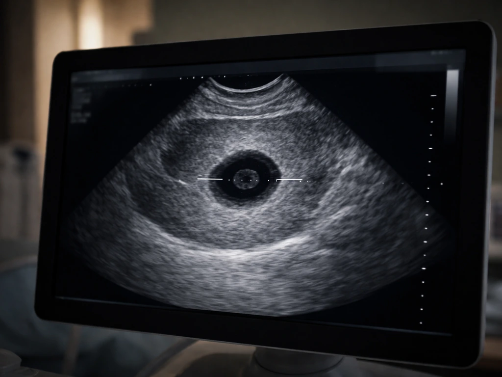
A gestational sac should generally become visible on transvaginal ultrasound once hCG reaches roughly 1,500 to 2,000 mIU/mL. With endometriosis, the key question is often where endo can grow and what signals and environment let it expand how endo grows. If the sac isn't visible or isn't growing when hCG is at that level or above, something has gone wrong with one of those inputs.
Physical space is the third requirement that often gets overlooked. A sac can only expand if its surrounding tissue is compliant enough to allow it. Scar tissue, a thick cyst wall, mechanical compression from adjacent structures, or abnormal internal pressure can all cap growth even when cellular machinery is functioning normally. Think of trying to inflate a balloon inside a cardboard box: the machinery works, but the environment won't allow expansion.
Why a sac stalls: the common culprits
Nutrient and signal shortfalls
For a gestational sac, inadequate hCG production is usually the first signal that something is wrong. A normally progressing early pregnancy roughly doubles hCG every 48 to 72 hours. Slow or plateauing hCG often precedes a sac that stops growing on ultrasound. For fluid-filled cysts and follicles, the equivalent failure is a drop in the hormonal or molecular signals (like FSH for ovarian follicles, or cAMP-linked pathways for epithelial cysts) that would otherwise drive both cell division and fluid secretion.
Transport limits and poor vascularization
Even perfect signals can't help if nutrients and oxygen can't get delivered. In follicles, for example, the fluid inside comes partly from thecal capillaries and partly from secretion by granulosa cells. Impair that blood supply, and fluid accumulation slows. The same principle applies to any sac that depends on a vascular bed: poor capillary development or disrupted Starling forces (the pressure dynamics that govern fluid movement across vessel walls) will starve the sac of the raw material it needs to grow. Lymphatic drainage problems can also reverse fluid accumulation, essentially draining the sac faster than it fills.
Ion channel and osmotic failure
A cyst or vesicle fills with fluid because epithelial cells actively pump ions (particularly chloride, powered by Na+/K+-ATPase activity) to create an osmotic gradient. Water follows. If those ion channels are disrupted, blocked, or genetically defective (as in some forms of polycystic kidney disease), the osmotic engine stalls and the sac can't inflate even if the cells are dividing normally. This is why some cysts grow aggressively in certain genetic conditions: the ion pump is overactive. A stalled cyst often has the opposite problem.
Cell-level failures: when the machinery breaks down
Growth at the cellular level means mitosis, differentiation, and programmed developmental steps happening in the right order. For a polymer that grows to form a polypeptide, the process depends on the ribosome, where amino acids are assembled into a chain. For a gestational sac, the embryo needs to implant successfully, develop a yolk sac, and then begin forming an embryonic pole with detectable cardiac activity.
When this sequence breaks down, you get what clinicians call a 'blighted ovum' or anembryonic pregnancy: a gestational sac that forms but contains no viable embryo. On transvaginal ultrasound, [the diagnosis can be confirmed when the mean sac diameter (MSD) reaches 25 mm or more with no embryo visible. ](https://www. acog.
org/clinical/clinical-guidance/practice-bulletin/articles/2018/11/early-pregnancy-loss) A sac that measures 21 mm or more with no embryo and no yolk sac is also highly suspicious for pregnancy failure.
For other types of sacs, impaired mitosis means the epithelial or mesothelial cells lining the sac wall stop dividing. This can happen because of DNA damage, p53-pathway activation, nutrient starvation, or simply because the developmental program that was supposed to drive growth reaches its endpoint. Programmed growth arrest is actually normal in many biological structures: an ovarian follicle that doesn't get the right hormonal surge at the right time will stop growing and undergo atresia (programmed regression). Growth not happening isn't always a malfunction; sometimes it's the system working exactly as designed.
Physical constraints that cap expansion
Even a biologically healthy sac hits physical limits. Internal pressure builds as fluid accumulates, and at some point that pressure equals the osmotic driving force pushing water in. Net fluid flow stops. This is a mechanical equilibrium, not a biological failure. In synovial joints, for instance, small subatmospheric pressures and a 'trans-synovial pump' mechanism regulate fluid volume actively, preventing runaway expansion. Your body has multiple feedback loops designed to prevent any fluid-filled space from growing indefinitely.
Structural defects in the sac wall itself can also prevent growth. If the wall is too rigid, too fibrotic, or if there's external compression from neighboring tissue, the sac simply can't expand mechanically even if it has all the biological tools to do so. Diffusion limits play a role too: the center of any sac that grows beyond a few millimeters needs a dedicated supply chain (blood vessels) to stay viable. Without vascularization, the core becomes hypoxic, cells die, and growth stalls. This is exactly the same diffusion-distance limit that constrains how large a single cell can grow before it needs to divide.
How to troubleshoot today
If it's a gestational sac
- Note the exact gestational age (based on last menstrual period or IVF transfer date) and the mean sac diameter measured on your ultrasound report. These two numbers together are the foundation of interpretation.
- Check whether your hCG was measured alongside the ultrasound. If hCG is above 1,500 to 2,000 mIU/mL and no intrauterine sac is visible, that needs same-day clinical attention.
- Ask whether a repeat ultrasound was scheduled. Single early scans are often inconclusive, and guidelines generally recommend a repeat 1 week later to confirm a diagnosis of pregnancy failure rather than acting on one scan alone.
- Document any symptoms: pelvic pain (especially one-sided), vaginal bleeding, or shoulder-tip pain. These change the urgency level significantly (see the red flags section below).
- Ask your provider specifically whether the sac location has been confirmed inside the uterus, because the entire risk profile changes if it hasn't been.
If it's a fluid-filled cyst, follicle, or other biological sac
- Record the context: is this being observed on serial imaging (e.g., ovarian follicle tracking) or is it a known cyst being monitored? What baseline size was documented and over what time period?
- Consider the signaling environment: for ovarian follicles, where are you in the menstrual cycle? Follicular growth is tightly tied to FSH and LH timing. A follicle that 'stops growing' mid-cycle may simply be waiting for its hormonal cue.
- For lab or experimental sacs/organoids: check ion transport conditions, osmolarity of the growth medium, and whether the scaffold or surrounding matrix is mechanically allowing expansion.
- Note any signs of regression or reabsorption (decreasing size over serial measurements) versus simply plateaued growth, as these suggest different mechanisms.
Red flags: when to get help immediately
If your situation involves a gestational sac, some scenarios cannot wait for a follow-up appointment. Ectopic pregnancy is the main concern. An ectopic pregnancy grows in the wrong location (usually a fallopian tube), and because a tube can't expand the way a uterus can, it can rupture, causing life-threatening internal bleeding. The sac in an ectopic pregnancy may not be visible on ultrasound inside the uterus at all, which is classified as a 'pregnancy of unknown location' (PUL) until proven otherwise. What clinicians sometimes see instead is a pseudogestational sac inside the uterus that looks like a true gestational sac but isn't.
Go to an emergency department or call emergency services immediately if you are pregnant (or think you might be) and you have any of these symptoms:
- Sudden or severe one-sided pelvic or abdominal pain
- Shoulder-tip pain (especially on the right side when lying down), which can signal internal bleeding irritating the diaphragm
- Heavy vaginal bleeding with clots
- Dizziness, fainting, or feeling like you might collapse
- A positive pregnancy test with no intrauterine sac confirmed on ultrasound and hCG above 1,500 to 2,000 mIU/mL
For non-gestational sacs, urgent medical attention is needed if a cyst shows rapid enlargement, signs of infection (fever, localized pain), or if it's in an anatomical location where rupture or torsion (twisting) could compromise blood supply to an organ, like an ovarian cyst causing ovarian torsion.
The bigger picture: growth failure as a biological story
Whether you're looking at a gestational sac, an ovarian follicle, an epithelial cyst, or even a vesicle in a lab model, the reason growth stalls almost always traces back to the same short list: missing inputs, broken signaling, impaired cell division, inadequate transport, or a physical ceiling imposed by mechanics and pressure. Pulses, like other living structures, grow only when they have the right nutrients, signals, and physical space for expansion pulses grow. These aren't random failures. They're the predictable outcomes when the tightly coordinated system that drives biological growth loses one of its key components.
Understanding this is actually useful beyond the immediate problem. The same diffusion limits that stop a sac from growing past a certain size without vascularization are the same constraints that explain why cells can't grow indefinitely before dividing, why tumors need to recruit blood vessels to grow large, and why any biological structure eventually hits a ceiling.
That same idea connects to the question of what continues to grow after death, since growth typically stops once the inputs and coordination are gone why any biological structure eventually hits a ceiling. Growth isn't free or automatic; it's an active, resource-intensive process that requires continuous coordination between chemistry, physics, and genetics. When any one of those threads breaks, the structure stops expanding, and your job is to figure out which thread snapped.
| Sac Type | Primary Growth Drivers | Common Stall Causes | Key Next Step |
|---|---|---|---|
| Gestational sac | hCG signaling, implantation, embryo development | Anembryonic pregnancy, ectopic location, chromosomal failure | Serial hCG + repeat transvaginal ultrasound in 1 week (or ER if red flags) |
| Ovarian follicle | FSH, LH surge, granulosa cell secretion | Hormonal insufficiency, anovulation, cycle timing mismatch | Cycle-day tracking, hormonal panel with provider |
| Epithelial cyst | cAMP signaling, chloride/ion transport, cell proliferation | Ion channel disruption, osmotic failure, fibrotic wall | Serial imaging to document size trend; specialist review if symptomatic |
| Synovial/joint sac | Mechanical pumping, Starling forces, lymphatic drainage | Joint disease, impaired lymphatic return, fibrosis | Rheumatology or orthopedic assessment if causing symptoms |
| Experimental vesicle/organoid | Growth medium composition, scaffold compliance, ion balance | Osmolarity mismatch, matrix stiffness, signal absence | Check medium formulation, matrix mechanical properties, signaling components |
FAQ
If a gestational sac looks small on ultrasound, does that always mean the pregnancy is failing?
Not always. Measurements can vary by scanner settings, operator technique, and the exact timing of ovulation. What matters more than a single scan is whether hCG is rising appropriately and whether serial ultrasound shows consistent interval growth. If hCG is at or above expected visibility ranges but the sac is not progressing, clinicians escalate evaluation sooner.
How can ectopic pregnancy affect the “sac not growing” picture?
In ectopic pregnancy, a sac may not appear in the uterus at all, or a misleading intrauterine appearance can occur. This is why clinicians often use the term pregnancy of unknown location (PUL) when imaging and hCG trends do not fit an intrauterine pregnancy. Rising hCG without a normal intrauterine pattern triggers careful, time-sensitive follow-up.
What is the difference between a cyst that should regress and one that is truly concerning?
Many ovarian follicles or functional cysts naturally stop enlarging and regress when hormone signals change. Red flags that prompt urgent assessment include rapid growth, severe or focal pain, fever or signs of infection, and risk of torsion based on size and symptoms. Without symptoms, a watch-and-recheck plan is commonly used for stable-appearing cysts.
My cyst or follicle isn’t getting bigger, is that necessarily good news?
Often it is, but the context matters. A stable size can mean it is involuting, but it can also reflect a blocked process, persistent inflammation, or a non-functional lesion. Clinicians look at features on imaging (solid components, septations, vascularity) and symptoms to decide whether observation is enough or whether further testing is needed.
Could medication or supplements change why a sac is not growing?
Sometimes. Hormonal therapies that affect ovulation or early embryo support can alter hCG and signal pathways. Also, any medication that changes hydration, bleeding patterns, or infection risk can indirectly affect evaluation timing and symptom interpretation. The safest approach is to tell your clinician exactly what you are taking and when, so serial hCG and ultrasound timing can be interpreted correctly.
How do clinicians decide whether to repeat ultrasound versus check hCG again?
They use both the hCG level and the time since the last measurement or scan. When hCG is near expected thresholds for sac visibility, repeating hCG and repeating ultrasound at an appropriate interval helps distinguish slow viability from measurement timing. If symptoms suggest ectopic or rupture risk, the threshold for urgent repeat evaluation is much lower.
What can cause “no embryo visible” even when a gestational sac is present?
A sac can be present without an embryo due to impaired early developmental sequence or delayed dating relative to ovulation. Clinicians account for gestational timing and use additional criteria based on sac size and presence of yolk sac before diagnosing failure. If criteria are not met, repeat imaging may be recommended rather than assuming anembryonic pregnancy immediately.
Does a “blighted ovum” mean the sac stopped growing because of ion transport or signaling?
In practice, the underlying label describes the observed ultrasound outcome, not a single molecular cause. The problem could involve inadequate early signaling for implantation and developmental progression, inadequate embryo viability, or timing mismatch. The key distinction is diagnostic and management-based, clinicians focus on serial trends and definitive ultrasound criteria rather than guessing one mechanism.
For non-gestational sacs, when should someone seek emergency care?
If a cyst is rapidly enlarging, associated with severe pain (especially one-sided), vomiting, dizziness, fainting, fever, or suspected torsion or rupture, emergency evaluation is appropriate. Timing matters because torsion can compromise blood supply quickly. If symptoms are mild but persistent, urgent clinic evaluation is still advisable to set the right follow-up interval.
Can mechanical pressure alone stop growth, even if signals and nutrients seem normal?
Yes. Physical constraints like a rigid or fibrotic wall, external compression, or unfavorable compartment anatomy can cap expansion even when the “biological machinery” is working. That is one reason imaging interpretation matters, because two lesions with similar cell-type behavior can behave differently depending on surrounding tissue compliance.
What’s a common mistake when people try to interpret sac measurements at home?
Over-interpreting one measurement or comparing results taken at different gestational ages or with different scan types. A single scan can miss the true growth curve, especially if ovulation timing was uncertain. Clinically, serial assessment using defined intervals and consistent measurement approach is what reduces misclassification.
Next Article
What Site Does the Polypeptide Grow On in Translation
In translation, a polypeptide grows on the ribosome, from the P site via the peptidyl transferase center as tRNAs cycle.