
Yes, microtubules can technically add tubulin at both ends, but the two ends behave very differently. The plus end is the fast, dynamic end where most of the action happens: rapid growth, sudden collapse, and rescue. The minus end grows much more slowly, usually around 2 to 3 times slower than the plus end in vitro, and in living cells it is often capped or anchored at an organizing center so it barely grows at all. So when someone asks 'can microtubules grow from both ends,' the honest answer is: yes in principle, but in practice, the plus end dominates and the minus end is usually under tight biological control.
Can Microtubules Grow From Both Ends? Plus vs Minus Growth

Marcus Whitmore
17 May 2026
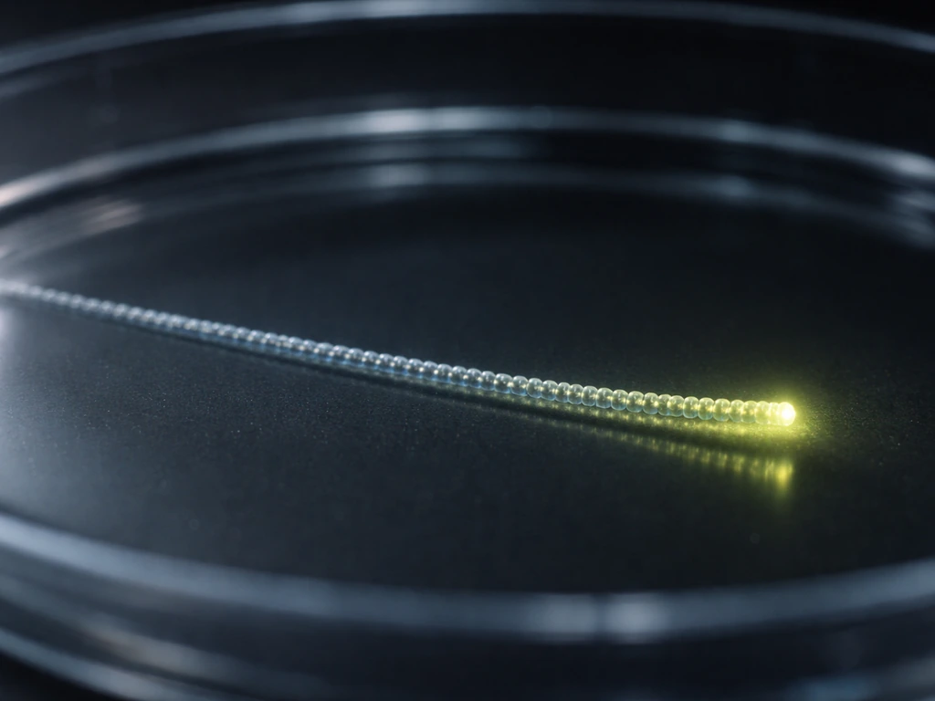
Microtubule ends: plus vs minus identity
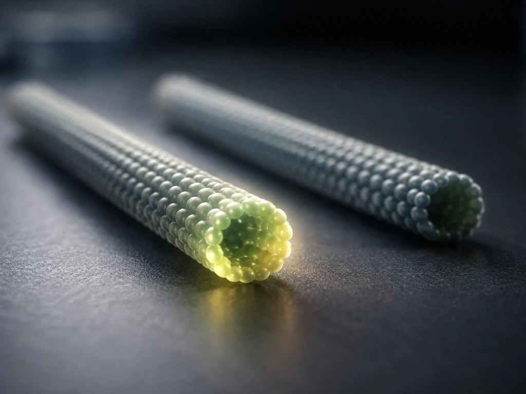
Every microtubule is a polar structure. It has a plus end and a minus end, and they are chemically different, not just labeling conventions. The plus end exposes beta-tubulin to the surrounding solution, while the minus end exposes alpha-tubulin. That structural difference is why the two ends behave differently in the first place: the geometry of the tubulin heterodimer means one end is inherently more permissive for adding new subunits quickly.
In most animal cells, microtubules originate at microtubule organizing centers (MTOCs) like the centrosome. The gamma-tubulin ring complex (gamma-TuRC) at the MTOC nucleates new microtubules and then caps the minus end, essentially locking it in place. The plus end then grows outward toward the cell periphery. That spatial arrangement, minus end anchored and plus end pointing outward, is the default setup you will encounter in most textbook diagrams and most standard cell biology experiments.
It is worth noting that this is not universal. Neurons, for example, have microtubules with mixed polarity in dendrites, and plant cells lack centrosomes entirely. Polarity still exists, but where the minus end ends up varies by cell type. Knowing the cell type you are studying is the first thing to clarify before making assumptions about which end is doing what.
What 'growing from both ends' really means
There is an important distinction between polymerization happening at an end and net elongation occurring there. Tubulin subunits can be added to the minus end in a test tube, and in vitro experiments using GMPCPP-stabilized microtubule seeds have directly measured growth rates at both ends under controlled conditions. So biochemically, both ends can polymerize. The question is whether they do so in a meaningful, sustained way inside a real cell, and for most cell types, the answer is mostly no for the minus end.
Think of it like a construction crew working on both ends of a tunnel. Both crews can technically dig, but one crew (the plus end) has three times the tools and no supervisor stopping them, while the other crew (the minus end) is working with fewer resources and often has a concrete plug (the gamma-TuRC cap) blocking their side. Net progress happens overwhelmingly at the plus end, even if a little activity is technically occurring on both sides.
Another source of confusion is nucleation versus elongation. Nucleation is the de novo creation of a new microtubule, which requires the gamma-TuRC template at the minus end. γ-TuRC structures have been reported as asymmetrically topped, capped ring-like templates that are consistent with oriented microtubule nucleation blank" rel="noopener noreferrer">gamma-TuRC template at the minus end. Elongation is the continued addition of tubulin to an existing microtubule, which is mainly a plus-end story in cells. Conflating these two processes leads to the mistaken idea that because the minus end is involved in nucleation, it must also be the main growth site. It is not.
Dynamic instability at each end
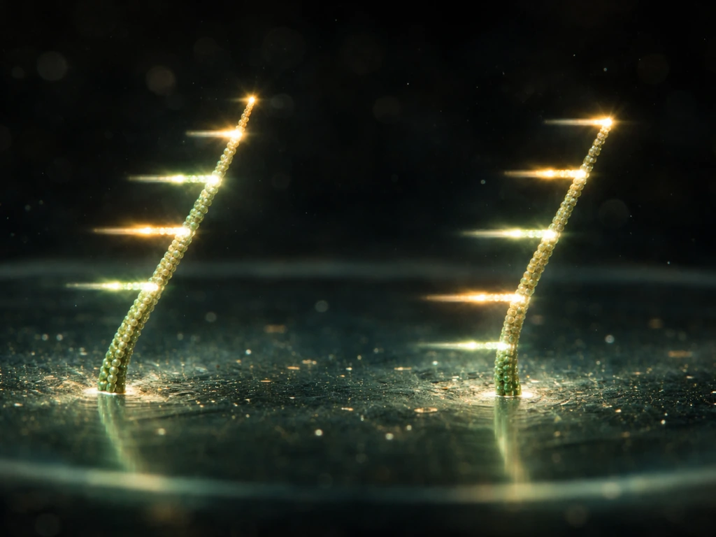
Dynamic instability is the defining feature of microtubule behavior: a single microtubule can rapidly switch between growing and shrinking without warning. This is driven by the GTP cap. Tubulin binds GTP when it joins the filament, and as long as the tip retains a cap of GTP-bound tubulin, the microtubule is stable and keeps growing. When that cap is lost because hydrolysis catches up with polymerization, the microtubule enters catastrophe: a rapid, uncontrolled depolymerization. Rescue is the reverse event, when a shrinking microtubule suddenly starts growing again.
At the plus end, these transitions are frequent, stochastic, and well-studied. Plus ends are the main venue for dynamic instability in most cells. The minus end also exhibits dynamic instability in vitro, but the rates and frequencies are quite different. Growth at the minus end is slower, catastrophe behavior is less dramatic, and in vivo the minus end is often stabilized by capping proteins or anchored entirely at the MTOC, so you rarely see classic dynamic instability behavior there unless you are working with free minus ends, which do occur in certain contexts like acentrosomal microtubules in neurons.
Experiments with drugs like nocodazole have confirmed that catastrophe and rescue processes exist at both ends, not just the plus. At nanomolar concentrations of nocodazole, researchers have observed decreases in elongation velocity, increases in catastrophe frequency, and decreases in rescue frequency at both plus and minus ends. This tells us the GTP-cap mechanism operates at both ends, just with different kinetics. Paclitaxel, on the other hand, suppresses shortening rates primarily by stabilizing the filament lattice, with measurable effects on plus-end mean squared displacement, confirming that the plus end remains the dominant dynamic endpoint.
What tips the balance: tubulin supply, proteins, drugs, and cell context
Several factors can shift how much growth actually occurs at each end, and understanding them helps explain why experimental results sometimes look like 'both ends growing.' In the same way that GTP-cap dynamics and end regulation control microtubule growth, plant and fungal systems also depend on signaling cues that what stimulates the pollen tube to grow, showing how context drives a directed growth response.
- Tubulin concentration: At very high free tubulin concentrations in vitro, both ends can show net polymerization simultaneously. Below a critical concentration, the minus end shrinks even while the plus end grows, a phenomenon called treadmilling.
- Plus-end tracking proteins (+TIPs): Proteins like EB1, CLIP-170, and XMAP215 accumulate at growing plus ends by recognizing the GTP cap or the geometric curvature at the tip. They promote plus-end growth and stability, amplifying the asymmetry between the two ends.
- Minus-end regulatory proteins: Proteins like Patronin and CAMSAP family members protect minus ends from depolymerization and can even promote minus-end growth in specific contexts, such as dendritic microtubule organization in neurons.
- Motor proteins: NuMA recruits dynein activity to microtubule minus ends during mitosis, and plus-end-directed kinesins preferentially associate with the plus end. These motor-end interactions reinforce the functional differences between the two ends.
- Severing enzymes: Spastin cuts microtubules and generates a new shrinking plus end and a nonshrinking minus end at the cut site. This asymmetric outcome after severing directly shows that the two newly created ends do not behave the same even moments after being generated.
- Drugs: Nocodazole destabilizes microtubules at both ends in a dose-dependent way. Paclitaxel kinetically stabilizes the lattice and suppresses plus-end shortening. These pharmacological tools let researchers dissect end-specific dynamics in experiments.
Cell context matters enormously. In a standard interphase fibroblast, minus ends are anchored at the centrosome and plus ends are free and dynamic. In a mitotic cell, both ends of spindle microtubules are under heavy regulation: kinetochore attachment happens at the plus end, while motor proteins at the spindle poles manage minus-end dynamics. In neurons, dendrites have microtubules with mixed orientation, meaning some microtubules present their minus ends toward the cell periphery, flipping the usual rule entirely.
How to interpret experiments and microscopy for end-specific behavior
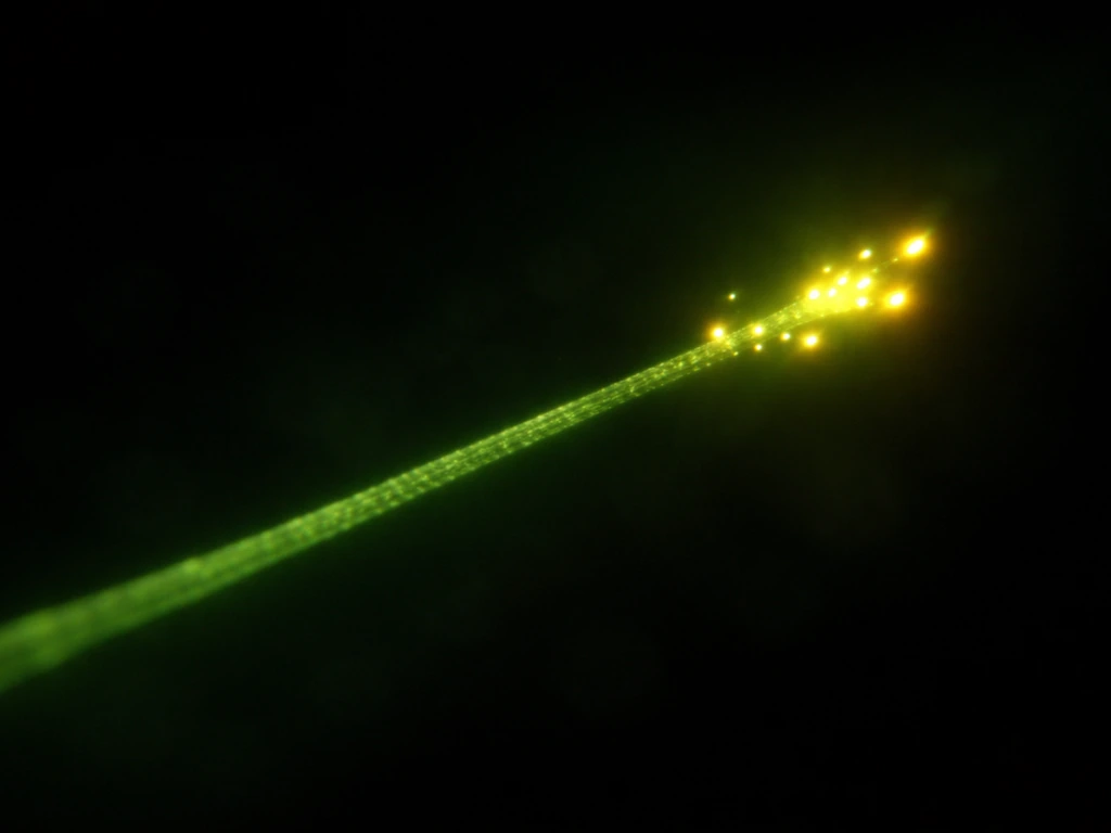
If you are looking at live-cell imaging or analyzing published data, knowing what to watch for at each end makes a huge practical difference.
Spotting plus-end dynamics
The gold-standard marker for growing plus ends is EB1 (or EB1-EGFP), which forms characteristic comet-shaped signals at the tips of polymerizing microtubules. The comet appears because EB1 binds the GTP-rich region near the tip and dissociates as GTP is hydrolyzed behind it. If you see comets, you are watching plus-end polymerization in real time. Kymographs, space-time plots generated from time-lapse images, let you measure growth rate, catastrophe frequency, and rescue frequency directly from the slope and shape of the trace. Comets disappearing abruptly on a kymograph signal a catastrophe; a sudden reappearance signals a rescue.
Catching minus-end behavior
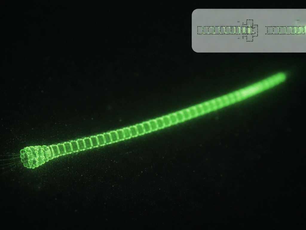
Minus-end dynamics are much harder to see in standard imaging because the minus end is usually anchored or capped and does not produce EB1 comets. To study minus ends experimentally, researchers often use one of three strategies: fluorescently labeled Patronin or CAMSAP proteins that mark minus ends specifically, acentrosomal microtubule preparations where both ends are free (sometimes generated by spastin severing or nocodazole washout), or permeabilized-cell reconstitution setups where drug exposure sequences let you separate plus-end from minus-end contributions. Using EB1 as a polarity marker in fixed cells, alongside a minus-end marker like NuMA, is a practical approach for determining which end is which in a static snapshot.
A quick comparison of what each end looks like experimentally
| Feature | Plus end | Minus end |
|---|---|---|
| Growth rate (in vitro) | Fast (~1–3 µm/min typical) | Slow (~0.5–1 µm/min, ~2–3x slower) |
| Dynamic instability in vivo | Frequent, well-documented | Rare in cells; present in vitro |
| GTP cap | Yes, drives catastrophe/rescue | Yes, but less studied in vivo |
| Common marker protein | EB1, CLIP-170, XMAP215 | Patronin, CAMSAP, NuMA (mitosis) |
| Typical cellular status | Free and dynamic | Anchored or capped at MTOC |
| Response to nocodazole | Increased catastrophe, decreased rescue | Same trends, different baseline rates |
| Response to paclitaxel | Suppressed shortening, stabilized tip | Less studied, lattice-level stabilization |
Common misconceptions and better mental models
A few wrong ideas about microtubule growth show up repeatedly, so it is worth naming them directly.
- Misconception: 'Both ends grow equally.' Reality: The plus end is 2 to 3 times faster in vitro, and in cells the minus end is often capped so it barely grows at all. Equal growth at both ends is not the default.
- Misconception: 'Microtubules always polymerize at both ends in cells.' Reality: In most cell types, the minus end is anchored at an MTOC and capped by gamma-TuRC. Active polymerization at the minus end is the exception, not the rule, and requires specific proteins like Patronin to enable it.
- Misconception: 'Nucleation and growth are the same thing.' Reality: Nucleation (building a new microtubule from scratch using gamma-TuRC at the minus end) is completely different from elongation (adding tubulin to an already existing filament, mainly at the plus end).
- Misconception: 'Dynamic instability only happens at the plus end.' Reality: The minus end can undergo dynamic instability in vitro and in some cellular contexts. It is less prominent and slower, but it is mechanistically possible.
- Misconception: 'After a microtubule is cut, both new ends behave the same.' Reality: Spastin severing experiments show the new plus end typically shrinks rapidly while the new minus end is more stable. End identity is immediate and asymmetric even at the moment of creation.
A mental model that works well: think of a microtubule like a ratchet strap. The plus end is the free end you are pulling out, full of tension and movement, alternating between extending and snapping back. The minus end is the anchored end locked into a mechanism at the wall. Most of the dynamic behavior you observe lives at the free end, while the anchored end just holds the system in place. That asymmetry is structural, biochemical, and maintained by a whole network of proteins on both sides.
Takeaways and next steps
Here is what to walk away with. In pollen tubes, a related problem is how the cytoskeleton supports directed growth down the style. Microtubules can add tubulin at both ends, but the plus end is faster, more dynamic, and the dominant site of growth and dynamic instability in living cells. The minus end is typically anchored, capped by gamma-TuRC, and slow.
The GTP cap is the molecular switch controlling growth and catastrophe at both ends, but its consequences are most visible and impactful at the plus end. Proteins like EB1 mark growing plus ends, while Patronin and CAMSAP protect minus ends. Drugs like nocodazole and paclitaxel affect dynamic instability parameters at both ends but with different mechanisms and end-specific kinetics. Severing creates two ends that immediately behave differently, reinforcing that end identity is a physical property, not just a label.
If you want to dig deeper, here are the most useful search terms and concepts to follow up on: dynamic instability, GTP cap model, plus-end tracking proteins (+TIPs), EB1 comet assay, gamma-TuRC nucleation, Patronin and minus-end protection, treadmilling, catastrophe and rescue rates, nocodazole dose-response dynamics, and kymograph analysis of microtubule ends. The reviews 'Tracking the ends: a dynamic protein network controls the fate of microtubule tips' and 'Control of microtubule organization and dynamics: two ends in the limelight' (both in Nature Reviews Molecular Cell Biology) are the best single sources if you want authoritative overviews of the full picture. For minus-end biology specifically, the Journal of Cell Science 'Microtubule minus-end regulation at a glance' is a concise and well-organized entry point.
If you are working through a course or lab context, the biggest practical skill to build is being able to look at an EB1-comet image or kymograph and correctly identify which end is which, what growth and catastrophe events look like, and what the absence of EB1 signal at the other end implies about minus-end status. That ability to read the experimental readout is what separates rote memorization from real understanding of how microtubules grow.
Exploring how microtubules grow and shrink in more detail, including the biophysics of treadmilling and dynamic instability, is a natural next step from here. To focus on the mechanism, read about how do microtubules grow at the level of end identity, tubulin addition, and the GTP cap. In plant and animal systems, experimenters can measure this by tracking how the plus end extends over time while the minus end remains comparatively restrained predict how the coleoptile will grow.
FAQ
If microtubules can grow at both ends, why do researchers usually say growth is mostly at the plus end?
Because net elongation in cells is dominated by the plus end’s higher effective polymerization rate and more visible dynamic instability, while the minus end is commonly anchored or capped at the MTOC. Even when polymerization events occur at the minus end, they are often too slow or too constrained to contribute much to length changes over time.
Does “growing from both ends” mean microtubules elongate symmetrically at any given moment?
Not usually. The phrase can mix up two ideas: local tubulin addition can happen at both ends, but the microtubule’s length change depends on which end is achieving net growth. If one end is repeatedly losing subunits through slower or weaker dynamic cycles, overall length still increases mainly where growth outpaces shrinkage.
Can treadmilling happen if the minus end is capped or anchored?
Yes, but it depends on how you define the phenomenon. In many systems, treadmilling reflects preferential plus-end growth combined with turnover near the minus end, not equal activity at both ends. If the minus end is strongly stabilized, turnover may be limited and the observed behavior will shift from classic treadmilling toward mostly plus-end dynamics.
How can I tell whether the minus end is truly polymerizing versus just unanchoring or being reconfigured by cell mechanics?
Watch the minus end’s position relative to a known anchor (for example, centrosome or a minus-end marker) over time. If the apparent “growth” comes from the whole microtubule sliding, severing, or reorientation, the minus-end tip may not be adding subunits at a high rate. Using a minus-end-specific protein marker together with plus-end tracking helps separate biochemical growth from mechanical changes.
Why do EB1 comets sometimes disappear even if the microtubule remains stable?
EB1 reporting is tied to the presence and extent of a GTP-rich region near the tip. A microtubule can still be polymerization-competent or paused in a way that reduces the GTP cap signal without causing an immediate catastrophe. This is why kymographs and additional readouts (like tip tracking or drug perturbations) are often used alongside EB1.
What happens to both ends after severing by spastin or similar mechanisms?
Severing creates new ends that immediately adopt end-specific behaviors based on their structural identity. Typically, the newly created plus ends behave like plus ends (more dynamic), while the minus ends, although newly exposed, often become quickly regulated by minus-end protection systems. This rapid re-establishment of polarity is why severing can make it look like “both ends” are active, without implying that identity has been erased.
In vitro, can minus-end growth become comparable to plus-end growth?
It can become more comparable under conditions where minus ends are not strongly capped or anchored and where the experimental setup supports tubulin addition at both ends. For example, controlled seed experiments and conditions that adjust end exposure can reveal measurable minus-end polymerization, but in many standardized cell-like conditions the plus end still dominates net elongation.
Do drugs that affect microtubule dynamics change plus and minus ends in the same way?
No. The mechanisms differ, and that changes what you observe at each end. Nocodazole primarily shifts dynamic instability parameters in a way that affects both ends, while paclitaxel stabilizes the lattice and often alters plus-end behavior in ways consistent with reduced shortening and altered tip motion, with different kinetics than at the minus end.
What’s the most common mistake when interpreting “both ends growing” in live-cell imaging?
Assuming EB1 signals at the tip automatically mean that end is the plus end. EB1 marks growing plus ends, but misassignment of polarity can happen if microtubules are rearranged, severed, or oriented differently than expected. Pairing EB1 with a minus-end marker (for fixed cells) or using known polarity cues in the preparation reduces this error.
If I want to experimentally measure minus-end dynamics, what should I optimize first?
End identification and free versus anchored status. Minus-end behavior is hard to interpret when the tip is capped, anchored, or rapidly reorganized, because the biological system can mask polymerization. Choosing a preparation that frees minus ends (or deliberately labels them and controls attachment) is usually the first decisive step.
Next Article
What Weaves Webs as They Grow? Spiders, Silk, and Limits
Explains what weaves webs as they grow, how spider silk glands mature, and limits from energy and environment.