Multicellular organisms grow by doing three things at once: adding new cells through division, enlarging those cells, and organizing them into the right structures in the right places. It starts with a single fertilized egg, and every step after that is driven by tightly coordinated cell division, chemical signaling, and gene regulation that tells each cell what to become, when to divide, and when to stop.
How Do Organisms With Many Cells Grow and Develop
Marcus Whitmore
19 May 2026
What 'growth' actually means for a multicellular body
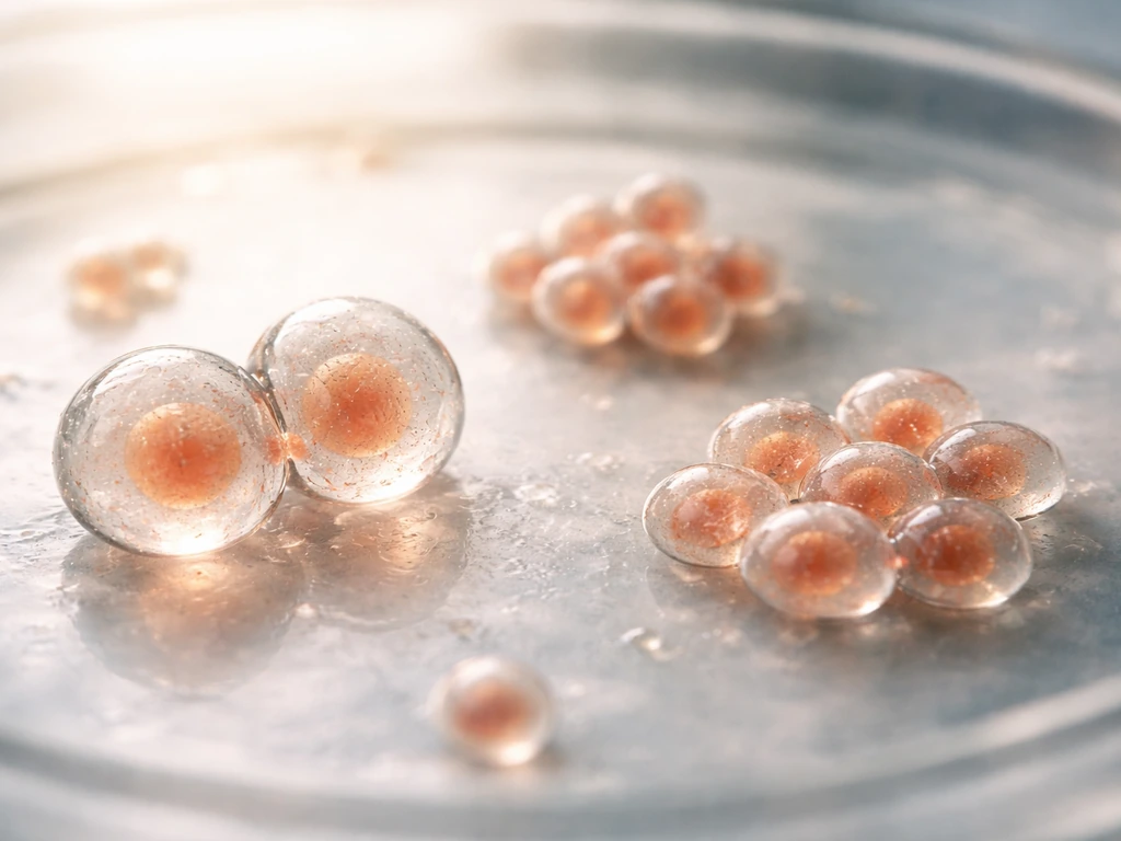
When we say a human or a dog or a tree 'grows,' we mean something more specific than just getting bigger. For a clear view of how organisms grow bigger, start by tracking both cell number increases and cell size expansion. Multicellular growth has three distinct components: increasing cell number (more cells through division), increasing cell size (each cell taking up more physical space), and building organized structure (cells arranging into tissues, organs, and body plans). A growing embryo is not just a pile of dividing cells getting larger. It is a system that simultaneously builds complexity. Miss any one of those three components and you don't get a functional organism, you get a disorganized mass.
This is fundamentally different from how single-celled organisms grow, where 'growth' mostly means one cell getting bigger until it divides into two. In a multicellular body, the cells that result from division don't just separate and go live independent lives. They stay connected, communicate, specialize, and contribute to a larger structure. That coordination is the whole game.
From one cell to many: division, mitosis, and differentiation
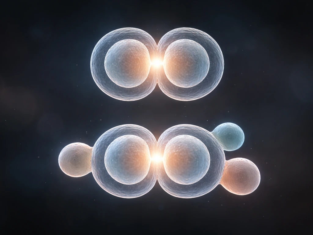
It all starts with mitosis. When a fertilized egg divides, it uses mitosis to produce two genetically identical daughter cells, and those cells do the same, and so on. Mitosis is the engine of cell number increase. Each round of division copies the cell's DNA completely, divides the chromosomes precisely, and splits the cell into two. Get that process wrong and you risk mutations, developmental errors, or uncontrolled growth.
But here's what makes multicellular growth remarkable: the daughter cells don't all stay identical. After enough rounds of division, cells begin differentiating, switching on different sets of genes depending on their position, the signals they receive, and their developmental history. A cell that ends up near the center of an embryo might become a muscle cell. One near the surface might become skin. Differentiation is how one genome produces hundreds of specialized cell types.
The decision to divide is itself regulated at specific checkpoints in the cell cycle. At the G1 checkpoint, a cell checks its size, its nutrient availability, growth factor signals, and whether its DNA is intact before committing to replicate. If growth factors are absent, Cdk inhibitors stay active in G1 and the cycle arrests. There's also a G2 checkpoint that blocks entry into mitosis if DNA replication is incomplete. These checkpoints mean division only happens when conditions are genuinely ready, not just because a cell could technically divide.
Building tissues and organs: signals, patterns, and coordination
Cell division and differentiation don't happen randomly. They are spatially and temporally coordinated by signaling pathways that act like a construction crew's blueprints. Signaling molecules diffuse across tissues, creating concentration gradients that cells use to figure out their position. Depending on how much of a signal a cell receives, it switches on a different gene program. This is how an embryo builds a body plan from scratch.
A great example is limb development. In a growing limb bud, mesenchyme cells proliferate to create the initial bud, and the overlying ectoderm is induced to form a structure called the apical ectodermal ridge (AER). FGF8 secreted by the AER feeds back to maintain mitotic activity in the mesenchyme below, driving outgrowth from shoulder to fingertip. FGF and Wnt signals work together to keep limb progenitor cells proliferating while staying undifferentiated, but also separately influence which cell lineages those progenitors will ultimately produce. It's a beautifully precise push-and-pull.
The formation of the vertebrate body's segmented structure (like your vertebrae) follows a 'clock-and-wavefront' mechanism. A molecular clock drives oscillating waves of Notch, Wnt, and FGF signaling across presomitic mesoderm cells. Notch signaling synchronizes these oscillations across neighboring cells so the clock ticks in unison. Where the wave meets a slowing wavefront of FGF activity, a new somite (the tissue block that will form a vertebra and associated muscles) is pinched off. The result is a rhythmically segmented body axis built with molecular-clock precision.
Organs follow the same logic of reciprocal signaling between neighboring tissue layers. In the developing lung, FGF10 secreted by mesenchyme drives repeated branching of the epithelial tubes, producing the tree-like airway architecture. The epithelial cells branch toward FGF10 signals, and as they do, they reshape the local signaling environment for the next branch. Growth and architecture emerge together.
Death is also part of building. Programmed cell death (apoptosis) sculpts tissues by eliminating specific cell populations at precise times. The spaces between your fingers formed because cells there died on schedule during embryonic development. BMP signaling can trigger this kind of death, and it is as essential to the final shape as any cell division.
Energy, nutrients, and transport: how cells get what they need
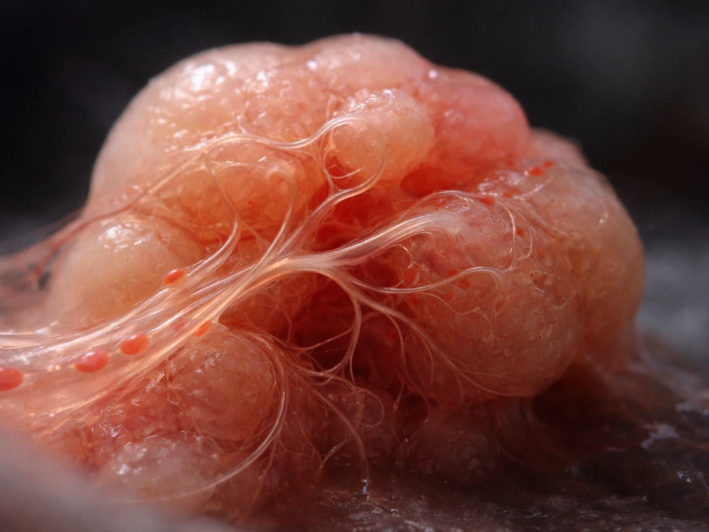
Every dividing cell needs raw materials: oxygen, glucose, amino acids, and the various molecular building blocks for new proteins, membranes, and DNA. In very small embryos, these can diffuse in directly from surrounding fluid. But diffusion has hard physical limits. Oxygen can diffuse effectively only about 100 to 150 micrometers through tissue before its concentration drops too low to sustain cell metabolism. That's roughly the width of a few human hairs.
This is why growing animals develop vascular systems. Blood vessels bring oxygen and nutrients right to the neighborhood of every cell, solving the diffusion bottleneck. As organisms get larger, the surface-area-to-volume ratio of any given tissue region drops, making passive diffusion even less effective. The evolutionary answer is always the same: specialized transport infrastructure. Without it, interior cells starve, and growth stops or goes wrong.
This constraint shapes growth strategy in practical ways. Tumors, for example, cannot grow beyond about 1 to 2 millimeters in diameter without recruiting new blood vessels (a process called angiogenesis). The same principle applies to normal developmental growth: tissues grow in coordination with the vascular networks that supply them.
Why growth isn't unlimited: checkpoints, contact inhibition, and programmed stops
Multicellular organisms don't just grow until they run out of food. Growth is actively stopped by multiple overlapping systems. The cell-cycle checkpoints at G1 and G2 are the first line of defense, blocking division when DNA is damaged or nutrients are insufficient. But developmental growth also has programmed endpoints built into it, controlled by timing genes and hormone signals that change at specific life stages.
Contact inhibition is a particularly elegant stop mechanism. When epithelial cells pack together tightly enough that they are touching neighbors on all sides, they arrest their own proliferation. This density-dependent growth arrest depends on intact cell-cell contacts and mechanical signals from the extracellular matrix. When you lose contact inhibition, as happens in cancer cells, proliferation continues past the point where it should stop.
Cellular senescence is another stop pathway. Cells that have divided many times, or that carry persistent DNA damage, activate the p53/p21 or p16/Rb pathways and enter a stable, irreversible cell-cycle arrest. This is a backup system: when checkpoints fail to catch a problem, senescence can still pull the brake. It's one reason why most DNA-damaged cells don't become cancerous, they just stop dividing permanently.
Together, these systems mean that growth in a healthy multicellular organism is always bounded: by signal availability, by physical contact, by developmental timing programs, and by surveillance mechanisms watching for errors.
Hormones, genes, and timing: who controls the growth schedule

Multicellular organisms don't grow at a constant rate throughout their lives. Growth is scheduled. The timing and rate are governed by hormones, gene regulatory networks, and environmental inputs that interact with the core cell-cycle machinery.
Insulin-like growth factor 1 (IGF-1) is one of the key regulators in animals. It promotes both proliferation and differentiation, and its effects on the cell cycle require continuous signaling through G1: if IGF-1 is present consistently, cells get the sustained push needed to pass the G1 restriction point and commit to division. Growth hormone works partly by stimulating IGF-1 production, linking the brain's assessment of nutritional status and developmental stage to tissue-level growth rates.
Gene regulatory networks set the developmental context in which these hormonal signals act. Transcription factors turned on at specific embryonic stages change which genes are responsive to growth signals, which is why the same hormone can have very different effects in a fetus versus an adult. Environmental inputs like nutrition, temperature, and stress feed into these regulatory networks too, which is why severe malnutrition during critical developmental windows causes permanent growth impairment rather than just temporary slowdown.
In plants, the equivalent hormonal regulators include auxin and brassinosteroids. Auxin drives cell elongation by regulating gene expression in the cell wall. Brassinosteroids control both cell division and cell expansion, and also regulate vascular differentiation. The principle is the same as in animals: chemical signals translate developmental and environmental information into regulated growth at the cellular level.
When growth goes wrong: disorders that reveal the mechanisms
Growth disorders are not just medical problems. They are also some of the clearest evidence for how the normal growth mechanisms work. When a control system fails, you can often figure out exactly what it was doing by seeing what goes wrong without it.
Cancer is the most vivid example of uncontrolled multicellular growth. Cancer cells typically have mutations in checkpoint proteins (p53 is mutated in roughly half of all human cancers), loss of contact inhibition, resistance to apoptosis signals, and the ability to recruit their own blood supply. Each of those features corresponds directly to one of the normal growth-limiting systems described above. Cancer doesn't invent new biology; it dismantles the controls that were already there.
Growth hormone deficiency in children produces proportional short stature because the IGF-1 signaling needed to sustain normal bone and tissue growth is insufficient. Conversely, excess growth hormone before growth plates close causes gigantism. Both conditions show how tightly hormonal signal levels are calibrated to produce normal growth trajectories.
Developmental patterning failures show the signaling side. Disruptions to Wnt, Notch, or FGF pathways during embryogenesis produce structural malformations: missing limb segments, vertebral fusion abnormalities, or branching defects in lungs and kidneys. These are not random failures. They map predictably onto the signaling events that those pathways control, confirming the mechanistic story.
Even senescence gone wrong matters. When cells bypass senescence without triggering full malignancy, they can remain metabolically active and secrete inflammatory signals that damage surrounding tissue, contributing to aging-related tissue dysfunction. The growth arrest that seemed like just a stop signal turns out to have broader consequences when it is leaky or incomplete.
Multicellular vs. single-cell growth, and how plants compare
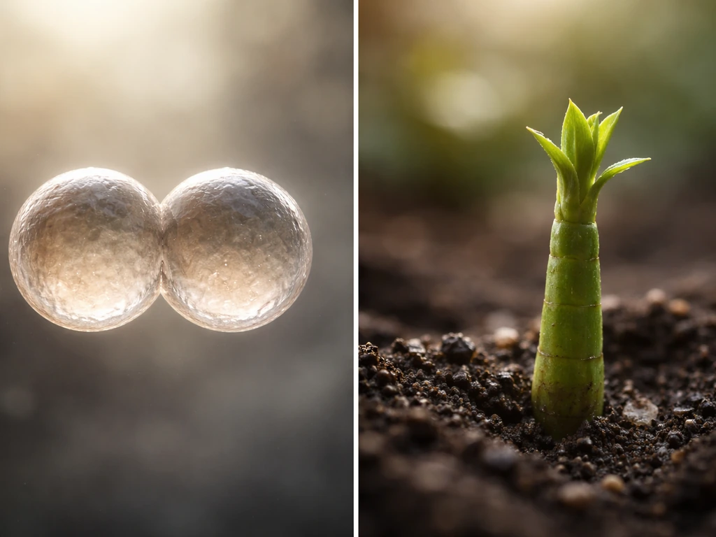
Understanding multicellular growth is sharpened by comparing it to simpler growth modes. The contrasts are instructive.
| Feature | Single-celled organisms | Multicellular animals | Multicellular plants |
|---|---|---|---|
| Primary growth mode | Cell enlargement then binary fission | Cell division (mitosis) plus differentiation | Cell division at meristems plus cell expansion |
| Coordination required | None (each cell is independent) | Extensive: signaling, patterning, checkpoints | Moderate: hormones, meristem organization |
| Differentiation | Absent or minimal | Hundreds of specialized cell types | Many specialized cell types, ongoing at meristems |
| Transport systems | Diffusion sufficient at small scale | Vascular systems (blood, lymph) required | Vascular tissue (xylem, phloem) required |
| Growth limits | Nutrient and surface-area-to-volume constraints | Checkpoints, contact inhibition, hormonal timing, diffusion limits | Meristem activity, hormones, resource availability |
| Growth timing | Continuous while nutrients allow | Developmentally programmed, stops at maturity | Often continuous (indeterminate) via persistent meristems |
Single-celled organisms do grow, but their growth is a prelude to division and essentially a solo act. There is no coordination with neighbors, no differentiation, and no tissue architecture to build. The surface-area-to-volume problem is real for single cells too (it sets an upper size limit, which is why most bacteria stay very small), but multicellular organisms solved it by organizing many small cells into systems with dedicated transport. That's explored in more detail in the context of how big single-celled organisms can actually get and what stops them.
Plants are an interesting middle case. They use the same core machinery (mitosis, hormonal regulation, signaling) but their growth is largely indeterminate: most plants keep growing throughout their lives because they maintain populations of undifferentiated stem cells in meristems at root tips and shoot tips. An animal's body plan is essentially fixed once development is complete; a tree keeps adding new rings, new branches, new roots. That ongoing growth relies heavily on cell expansion driven by water uptake and cell-wall loosening regulated by hormones like auxin, not just division.
The growth pipeline: a step-by-step summary
- A fertilized egg (zygote) begins dividing by mitosis, copying its genome faithfully and splitting into daughter cells.
- Early cell divisions produce a ball of cells; positional signals begin establishing which cells are 'inside' vs. 'outside,' 'top' vs. 'bottom.'
- Signaling gradients (Wnt, FGF, Notch, BMP, and others) create spatial information that cells use to switch on specific gene programs and begin differentiating.
- Differentiated cells organize into tissue layers and organs through reciprocal signaling between adjacent cell populations (epithelial-mesenchymal interactions, branching morphogenesis).
- Vascular systems develop in parallel with tissue growth, ensuring no cell ends up more than ~100 micrometers from an oxygen and nutrient supply.
- Cell-cycle checkpoints (G1 and G2 primarily) continuously gate division based on growth factor availability, nutrient status, cell size, and DNA integrity.
- Contact inhibition, apoptosis, and cellular senescence operate as additional stop signals to prevent overgrowth and eliminate damaged or surplus cells.
- Hormones (IGF-1, growth hormone, sex steroids in animals; auxin, brassinosteroids in plants) and gene regulatory networks set the pace and schedule of growth across developmental time.
- At developmental endpoints, programmed changes in gene expression and hormonal environment wind down growth and lock most somatic cells into non-dividing states.
Where to go next if you want to go deeper
If the cell division side of this is still fuzzy, the clearest next move is to really nail how mitosis works step by step and understand why it produces genetically identical daughter cells. From there, the concept of how mitosis specifically drives size increases in multicellular organisms becomes much more concrete. Once you have that, looking at cellular division and what it actually increases in terms of organism size will tie the mechanism to observable outcomes.
For the signaling and patterning parts, the best conceptual tools are understanding morphogen gradients, the concept of induction (one tissue instructing a neighboring tissue to change fate), and the logic of the cell cycle checkpoints. These three ideas connect most of the developmental biology described in this article. After those, exploring the two main ways organisms grow (cell number vs. These two main ways organisms grow are increasing cell number through division and increasing cell size through cell expansion the two main ways organisms grow. cell size) will give you a framework for comparing growth strategies across very different organisms, from bacteria to whales.
The most practical takeaway is this: multicellular growth is not one process, it is a pipeline of coordinated processes, each dependent on the one before it. Division without differentiation gives you a tumor. Differentiation without signaling gives you the wrong structures in the wrong places. Signaling without transport limits gives you structures that cannot be sustained. Understanding growth means understanding how all of these work together, and how each control point keeps the whole system on track.
FAQ
Do multicellular organisms need to grow by adding new cells, or can they grow mainly by increasing cell size?
They can do either, depending on tissue and life stage. Many animal tissues grow substantially by cell number early on, but later growth often relies more on cell expansion. Also, some organs are limited by how much fully differentiated cells can enlarge without losing function, so the balance between division and expansion varies by tissue.
What happens if cell division is blocked in a developing embryo, can growth still proceed?
Often part of growth slows or stops because fewer cells are produced to build structures. However, some tissues can still expand for a time through cell enlargement, and signaling can sometimes compensate by redirecting fate decisions. In most cases, persistent division failure leads to malformed or undersized organs because architecture requires both adequate cell number and correct patterning signals.
Why do organisms sometimes produce extra cells during development instead of stopping exactly on time?
Because development uses safety margins and quality control. Checkpoints and programmed death remove cells that are in the wrong place, not the right type, or produced under suboptimal conditions. This helps ensure the final tissue geometry and proportions are correct even when early signaling or differentiation is noisy.
How does a cell know when to stop dividing, is it only genetic timing or also physical conditions?
It is both. Cells respond to internal checkpoints (DNA integrity and nutrient status) and also to external cues, including cell density, contact signals, and mechanical feedback from the extracellular matrix. For epithelial tissues, high local density and strong neighbor contact can trigger growth arrest even when nutrients are available.
Is diffusion always the limiting factor for multicellular growth, or can cells overcome it without blood vessels?
Diffusion is a key limit over short distances, but some organisms reduce the need for long-range transport by staying thin, using diffusion-friendly body plans, or creating specialized internal channels. In many animals, however, scaling up requires active transport infrastructure such as blood vessels, because diffusion alone cannot maintain oxygen and nutrient levels deep inside tissues.
Why can cancer tumors grow a little before recruiting blood vessels?
Early tumor growth can be supported by diffusion from nearby existing vessels, so cells near the periphery survive. As the mass enlarges, inner cells fall below metabolic needs, and growth selects for cells that can trigger angiogenesis to reestablish supply lines.
Do plants use the same stop mechanisms as animals, like senescence and contact inhibition?
Plants do have growth arrest and regulated developmental transitions, but the details differ. Contact inhibition is less central than in many animal epithelia, while hormone-driven growth programs, tissue differentiation states, and meristem maintenance control whether cell division continues. Plants can also compartmentalize and continuously replace growing tissues, which changes how “stopping” looks across the whole organism.
Can hormones always be treated as the main control knob for growth?
Not always. Hormones often regulate growth by tuning cell-cycle responsiveness to other inputs, but the outcome also depends on which genes and signaling pathways are active in that tissue at that developmental stage. The same hormone level can produce different growth responses because transcription factor networks change what the cells can do.
What role does programmed cell death play if development depends on building more cells?
Programmed cell death is part of shaping, not only pruning. Removing specific cell populations creates spaces, boundaries, and tissue geometry that division alone cannot achieve. It also eliminates cells with incorrect signaling outcomes, helping refine patterns into functional structures.
If mitosis produces genetically identical daughter cells, how do cells still become different types?
Genetically identical cells diverge because they receive different signals and occupy different positions, which changes which genes get turned on. Differentiation often depends on cumulative developmental history and the timing of morphogen exposure, so two descendants from the same genome can end up with different stable expression programs.
Are cell cycle checkpoints only relevant to preventing cancer, or do they matter for normal development too?
They matter for normal development. Checkpoints ensure cells do not proceed to division when DNA replication is incomplete or when conditions are unsuitable, which preserves tissue integrity and prevents developmental errors. Developmental timing can also be sensitive to checkpoint activity, so checkpoint defects can cause both abnormal growth and abnormal patterning.
What is the fastest way to predict which growth failure might occur if a signaling pathway is disrupted?
Map the pathway to its known function in patterning versus growth control. If a pathway mainly sets positional information, disruptions often yield structural defects (mispatterned tissues). If it mainly sustains proliferation or branching, defects more strongly affect size, outgrowth extent, or organ architecture. Checking whether the tissue relies on diffusion gradients or reciprocal signaling can also narrow the likely outcome.
Citations
No web sources were captured for this target set (only partial search results unrelated to this specific “three main components of multicellular growth” textbook definition were returned). Please confirm you want me to do fresh targeted searches for textbooks/standard references (e.g., Alberts, Gilbert, Campbell) for the exact “increase in cell number / increase in cell size / formation of organized structures” wording.
/
Progression into S phase can be delayed at the G1 stage to allow cell growth and to be regulated by extracellular signals (showing checkpoints link cell-cycle entry to developmental/size programs rather than division-only).
https://www.ncbi.nlm.nih.gov/books/NBK26856/
In the absence of mitogenic signals, Cdk inhibition in G1 is maintained, arresting the cell cycle (illustrating how controlled division depends on signaling context in multicellular embryos).
https://www.ncbi.nlm.nih.gov/books/NBK26877/
Cell-cycle checkpoints act at G1, G2, and during mitosis (spindle checkpoint) to prevent division under unfavorable conditions such as DNA damage or inadequate conditions/space.
https://www.khanacademy.org/science/ap-biology/cellular-molecular-biology/stem-cells-and-cancer/a/cell-cycle-checkpoints-article
Commitment decisions include a major regulatory control in G1 (restriction point/R-point); entry into mitosis depends on regulated regulatory events that reset each new replication competence state in G1 after mitosis.
https://www.ncbi.nlm.nih.gov/books/NBK6318/
G2 checkpoint delays entry into mitosis when DNA replication is incomplete (preventing progression into division until genome replication is complete).
https://www.ncbi.nlm.nih.gov/books/NBK26856/
At the G1 checkpoint, cells decide whether to proceed based on factors such as cell size, nutrients, growth factors, and DNA damage.
https://www.khanacademy.org/science/ap-biology/cellular-molecular-biology/stem-cells-and-cancer/a/cell-cycle-checkpoints-article
FGF and Wnt signals synergistically promote proliferation while maintaining limb progenitors in an undifferentiated multipotent state, but act separately to determine lineage specification during limb development.
https://pubmed.ncbi.nlm.nih.gov/18776145/
Extrinsic Wnt-responsive stem-cell fate choices (self-renew vs differentiate) are dictated by signals with short range of action, helping limit stem-cell number and coordinating differentiation with proliferation control.
https://wnt.stanford.edu/stem-cells-and-wnt-signaling
Somite patterning links signaling gradients to tissue-state transitions; for example, mesenchymal-to-epithelial transition in the presomitic mesoderm correlates with changes regulated by FGF signaling (including Snai gene downregulation).
https://pmc.ncbi.nlm.nih.gov/articles/PMC2828275/
Somitogenesis can be decomposed into modules including (i) segmentation clock dynamics, (ii) segmental determination, (iii) anteroposterior polarity patterning, and (iv) epithelial morphogenesis; clock-and-wave dynamics are central to generating the segmented body plan.
https://www.nature.com/articles/s41580-024-00709-z
Somites are formed as transient epithelial (epithelial ball-like) structures from presomitic mesoderm; Notch signaling contributes to rhythmic expression programs in the segmentation process.
https://www.ncbi.nlm.nih.gov/books/NBK53154/
Notch signaling helps synchronize oscillations across neighboring presomitic mesoderm cells, supporting a “clock-and-wavefront” model for periodic somite formation.
https://www.nature.com/articles/35044091
In limb development, mesenchyme proliferation creates a limb bud bulge under the epidermis; mesenchyme-secreted factors induce overlying ectoderm to form the apical ectodermal ridge (AER), with FGF10 suggested as an inducing factor.
https://www.ncbi.nlm.nih.gov/books/NBK10003/
Reciprocal signaling between the AER and limb mesenchyme includes FGF8 secreted by the AER that maintains mitotic activity in the progress zone mesenchyme to drive proximal-distal outgrowth.
https://www.ncbi.nlm.nih.gov/books/NBK10102/
FGF signaling (e.g., FGF10) drives branching morphogenesis in the developing mouse lung, illustrating epithelial–mesenchymal signaling that assembles organs.
https://www.ncbi.nlm.nih.gov/books/NBK53153/
Developmental apoptosis eliminates specific cell populations at specific embryonic stages to shape tissues; this is integrated with other differentiation signals (e.g., BMP signaling can reproduce death-signal-like activity in certain contexts).
https://www.nature.com/articles/4401455
A cited quantitative limit: the maximum diffusion distance of oxygen from blood vessels is about 100 μm, and a typical radius of ~100–150 μm corresponds to the maximal diffusion distance of oxygen in tissue (relevant to growth/vascularization limits).
https://pmc.ncbi.nlm.nih.gov/articles/PMC6371795/
Surface-area-to-volume related constraints can couple to division timing in biological systems (supporting the general idea that SA:V constrains growth and division coordination).
https://www.sciencedirect.com/science/article/pii/S0966842X18301021
As animal/cell size increases, surface-to-volume ratio changes so diffusion becomes less efficient; multicellular organisms respond by specialized tissues/systems to complete limited nutrient/waste transport tasks.
https://bio.libretexts.org/Bookshelves/Introductory_and_General_Biology/Book%3A_General_Biology_%28Boundless%29/33%3A_The_Animal_Body-_Basic_Form_and_Function/33.04%3A_Animal_Form_and_Function_-_Limiting_Effects_of_Diffusion_on_Size_and_Development
Epithelial contact inhibition: confluence-dependent arrest of cell proliferation occurs in epithelial sheets, and loss of contact inhibition is a hallmark of cancer overgrowth.
https://www.nature.com/articles/srep46326
Contact inhibition depends on cell–cell contacts and matrix/ECM mechanics; disrupting these can relax density-dependent growth arrest, providing a mechanistic stop-system failure route to abnormal growth.
https://pmc.ncbi.nlm.nih.gov/articles/PMC3065384/
Apoptosis during embryogenesis includes programmed cell death at specific sites and times; for maize embryo formation, programmed cell death is reported in specific morphological/molecular contexts.
https://pmc.ncbi.nlm.nih.gov/articles/PMC4240416/
Cellular senescence involves stable cell-cycle arrest mediated by p53/p21 or p16/Rb tumor-suppressor pathways (pathways commonly implicated in cancer when disrupted).
https://www.cellsignal.com/science-resources/overview-of-cellular-senescence
Senescence growth arrest can be triggered by persistent DNA damage response signals (intrinsic or external); p53 induces p21 to block CDKs, leading to Rb-mediated cell cycle exit.
https://pmc.ncbi.nlm.nih.gov/articles/PMC5873888/
IGF1 is involved in growth, development, differentiation, and metabolism; IGF/IGF1 signaling is linked to proliferation and differentiation control (with distinct actions from growth hormone).
https://www.nature.com/articles/ncpendmet0427
Continuous IGF stimulation in G1 is required for IGF-induced cell proliferation, illustrating how hormone-mediated timing (duration/continuity of signaling) drives growth rate.
https://pmc.ncbi.nlm.nih.gov/articles/PMC3436179/
Auxin and brassinosteroid promote cell expansion by regulating gene expression, connecting plant hormone signaling to transcriptional control of growth.
https://www.genomebiology.biomedcentral.com/articles/10.1186/gb-2004-5-11-245
Brassinosteroids play roles in diverse developmental processes; the review notes cell divisions and cell expansion are main factors controlling plant organ growth, and evidence supports roles in regulating these outputs (including vascular differentiation).
https://academic.oup.com/plcell/article/32/2/295/6099060
Brassinosteroids are reported to play roles in controlling cell expansion, cell division, and vascular differentiation, based on plant studies and treatment experiments.
https://pubmed.ncbi.nlm.nih.gov/32143305/
Segmentation clock oscillations involve cyclic expression networks across Notch/Wnt/FGF; loss or interference of one cyclic program can adversely affect oscillatory gene expression and somite formation.
https://www.nature.com/articles/ncomms1381
Cells use checkpoints to arrest division when DNA is damaged and when conditions are unfavorable (mechanistic rationale for how growth is controlled and why checkpoint defects can lead to cancer).
https://www.khanacademy.org/science/ap-biology/cellular-molecular-biology/stem-cells-and-cancer/a/cell-cycle-checkpoints-article
Next Article
How Big Can Single-Celled Organisms Grow? Size Limits Explained
Learn what limits cell size and the largest known single-celled organisms, plus how to estimate size using diffusion and