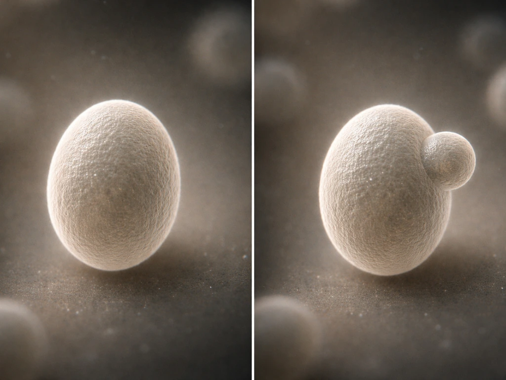
Cells grow and reproduce to maintain homeostasis by running a tightly regulated cycle: they build up biomass (proteins, lipids, organelles), check that everything is ready, copy their DNA exactly once, then split into two daughter cells. These steps also enable key organelles to grow and divide so the cell can reproduce itself copy their DNA exactly once, then split into two daughter cells. These same growth and division mechanisms show up in the cell cycle, which coordinates how cells grow and reproduce. The whole process is controlled by chemical signals that tell a cell when to divide, when to pause, and when to die, keeping tissue cell numbers and internal chemistry stable rather than spiraling into runaway growth.
How Do Cells Grow and Reproduce to Maintain Homeostasis
Marcus Whitmore
24 May 2026
Cell growth basics: what cells must build to get bigger
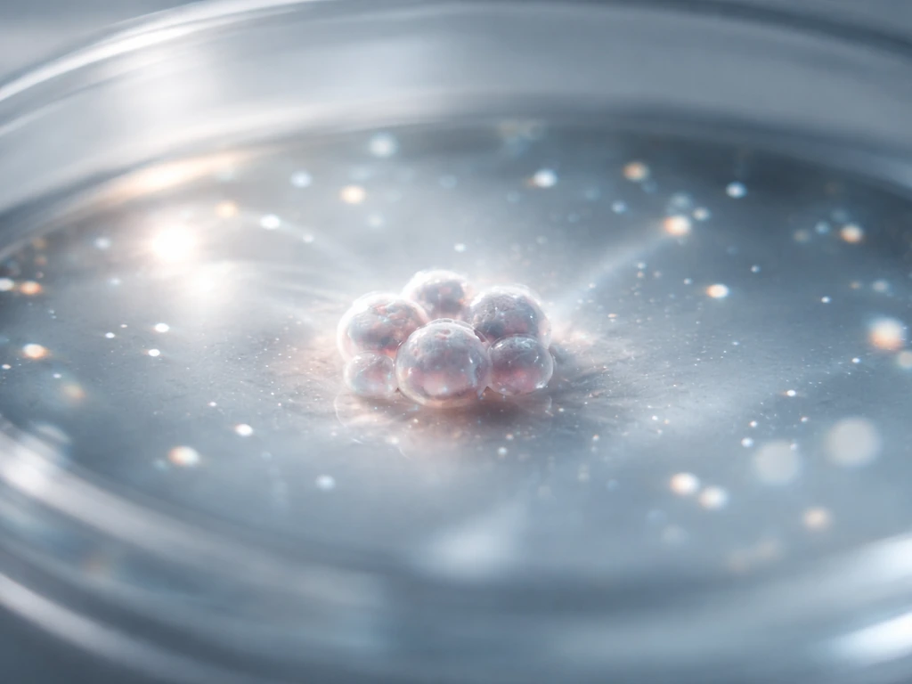
Before a cell can divide, it has to grow. Before human cells can grow and reproduce they need nutrients and energy to build the biomass for division. That means synthesizing enough protein to build new ribosomes, membranes, and enzymes; manufacturing lipids to expand the plasma membrane; duplicating organelles like mitochondria; and stockpiling the nucleotides needed for DNA replication. None of this is free. Growth is directly tied to nutrient availability and the cell's energy status.
Two master regulators sit at the center of this: mTORC1 and AMPK. Think of them as a gas pedal and a brake. When nutrients are plentiful, mTORC1 is active and phosphorylates targets that ramp up protein synthesis, mitochondrial biogenesis, and even nucleotide production through the oxidative pentose phosphate pathway. When energy runs low, AMPK flips the switch the other way, throttling anabolic processes and upregulating autophagy to recycle parts. A cell that tries to divide without enough raw material is a cell heading for trouble, which is exactly why these sensors exist.
Cell volume itself is also a homeostatic variable. Cells maintain a tightly defined intracellular ionic environment, using ATP-driven ion pumps and regulated water flow to resist osmotic stress. If a cell swells or shrinks too much, normal biochemistry breaks down. Volume regulation is not a side note; it is one of the earliest homeostatic acts a growing cell performs.
Homeostasis at cellular vs organism level
Here is a distinction worth locking in: homeostasis means something slightly different depending on the scale you are looking at. Inside a single cell, homeostasis is about chemical balance: stable pH, ion concentrations, redox state, and metabolite pools. A yeast cell living alone, for example, maintains these conditions entirely through its own sensors and transporters, with no neighbors to consult.
In a multicellular organism, homeostasis scales up to the tissue and organ level. Now the variables that need stabilizing include cell number, cell composition, extracellular matrix properties, interstitial fluid volume, oxygen and nutrient delivery, and metabolic waste removal. A gut lining that loses too many cells too fast stops functioning as a barrier. Skin that proliferates without limit becomes a tumor. Maintaining tissue homeostasis means coordinating cell birth (division) and cell death (apoptosis) so the net cell number stays in an appropriate range.
The practical implication: in multicellular organisms, individual cells must sometimes sacrifice their own interests, dividing when they get the signal and dying when they are no longer needed, in service of the organ's steady state. That cooperation is enforced by cell signaling, and when it breaks down, disease follows.
The cell cycle: checkpoints that synchronize growth and division
The cell cycle is the four-phase sequence every dividing eukaryotic cell runs through: G1 (gap and growth), S (DNA synthesis), G2 (second gap and preparation), and M (mitosis and division). It has both clock-like periodicity and switch-like checkpoints that prevent the cell from rushing through a phase before the previous one is finished.
Each checkpoint is enforced by a pairing of cyclin proteins with cyclin-dependent kinases (CDKs). In G1, cyclin D partners with CDK4 and CDK6 to push a cell toward commitment. At the G1/S transition, a cell reaches the restriction point, sometimes called Start, after which it is committed to dividing regardless of external signals. CDK2 with cyclin E drives this transition. Then CDK1 paired with cyclin B, a complex historically called MPF (maturation-promoting factor), is the master switch for entry into mitosis at the G2/M boundary.
Three major checkpoints act as quality-control gates. The G1/S checkpoint assesses whether the cell is big enough, nutrients are available, and DNA is undamaged. The G2/M checkpoint confirms that DNA replication is fully complete and any damage has been repaired before chromosomes are pulled apart. The spindle assembly checkpoint (SAC), operating inside mitosis itself, delays chromosome separation until every chromosome is correctly attached to spindle microtubules from opposite poles. SAC proteins Mad2, BubR1, and Bub3 inhibit the APC/C complex by blocking its co-activator Cdc20 until all kinetochores signal correct attachment.
A cell that fails a checkpoint does not just pause; it can be routed into permanent arrest or apoptosis. That is the safety net that keeps defective cells from reproducing. These processes enable a cell to grow and reproduce when conditions are appropriate can grow and reproduce.
How cells reproduce: DNA replication and mitosis/cytokinesis
Licensing and running DNA replication
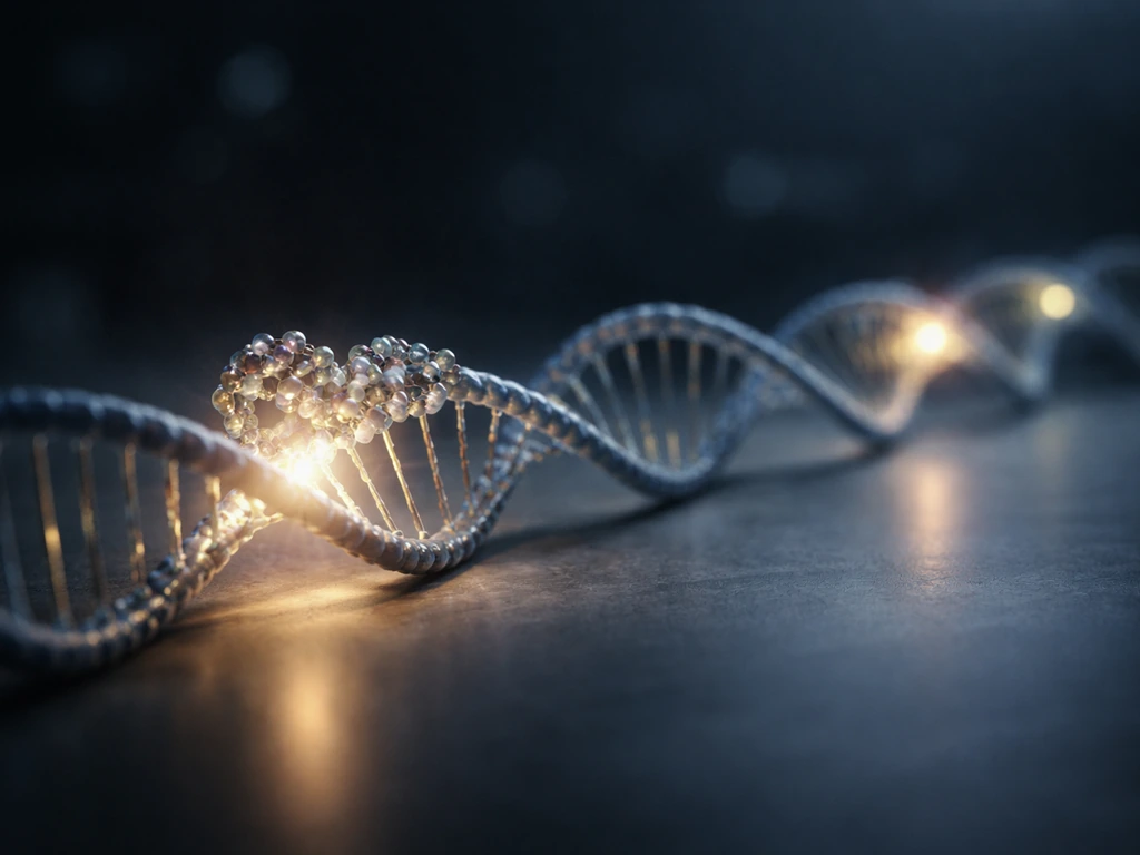
DNA replication follows a strict 'once and only once per cell cycle' rule. In late mitosis and early G1, before CDK activity rises high enough to fire origins, a six-subunit origin recognition complex (ORC, subunits Orc1 through Orc6) binds replication origins across the genome. ORC then recruits the MCM2-7 helicase complex to 'license' those sites for use. When S phase begins and CDK activity rises, licensed origins fire and replication proceeds. After an origin fires, high CDK activity prevents new MCM loading, so the genome cannot be re-replicated within the same cycle. PCNA, a ring-shaped sliding clamp, wraps around the DNA strand and keeps the polymerase machinery moving processively along the template.
Mitosis: segregating the chromosomes accurately
Once DNA is replicated, the two sister chromatids are held together by cohesin complexes. Cohesin is essential not just for keeping sisters paired but for ensuring they attach to microtubules from opposite spindle poles, a state called bi-orientation. At the metaphase-to-anaphase transition, the APC/C ubiquitin ligase complex (roughly 1.5 megadaltons in size) targets two key proteins for destruction: securin, which had been protecting the cohesin-cleaving enzyme separase, and cyclin B, whose destruction inactivates CDK1 and allows mitotic exit. Once securin is gone, separase cleaves cohesin, sister chromatids are pulled to opposite poles, and the cell commits to finishing division.
Cytokinesis: splitting the cell in two
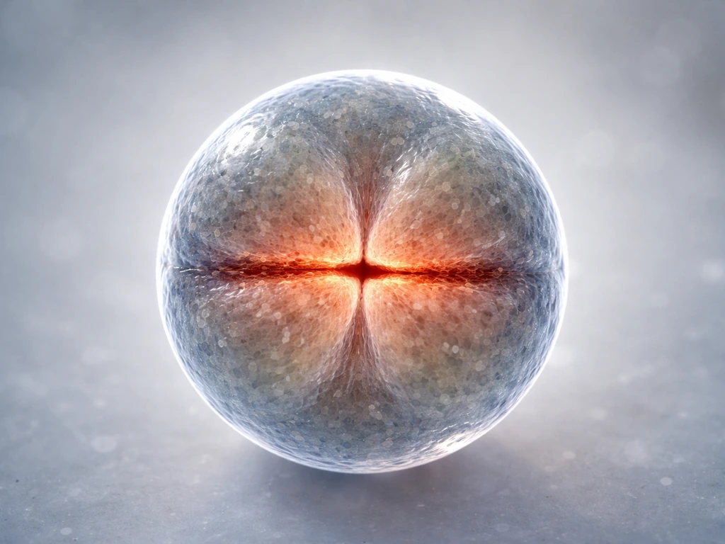
After chromosomes reach opposite poles, the cell physically divides by cytokinesis. In animal cells, a contractile ring of actin filaments and myosin-II assembles at the cell's equator. The GTPase RhoA drives ring assembly and activation. Polo-like kinases coordinate this with the mitotic spindle, ensuring the ring forms precisely where the two chromosome sets are separated. Master complexes called centralspindlin and the chromosomal passenger complex (CPC) regulate the final steps. The ring constricts like a drawstring until the two daughter cells pinch apart, each inheriting a complete genome and roughly equal cytoplasm.
How cell signaling controls when to divide or stop
No cell divides in isolation. In multicellular organisms, growth factors, cytokines, and cell-to-cell contact signals all feed into the decision to proliferate or hold. Growth factors bind surface receptors and activate intracellular cascades (like the RAS/MAPK and PI3K/AKT pathways) that ultimately upregulate the cyclins driving G1 progression. Remove the growth factor signal and most normal cells exit the cycle and sit in a quiescent state called G0.
Contact inhibition is one of the most important brakes on proliferation in tissues. When epithelial cells pack together, cadherin proteins on neighboring cells bind each other. This cadherin ligation activates the Hippo signaling pathway, which turns on LATS kinases that phosphorylate the transcriptional coactivators YAP and TAZ, excluding them from the nucleus.
Cadherin-mediated stimulation of Hippo signaling depends on cadherin ligation or homophilic bonding, which then drives LATS-mediated phosphorylation of YAP and its nuclear exclusion to connect cell-cell contact with growth inhibition cadherin ligation activates the Hippo signaling pathway. With YAP/TAZ sidelined, pro-growth gene expression drops.
Depletion of E-cadherin or its partner beta-catenin can release this brake and stimulate uncontrolled proliferation. In skin, proliferation is normally restricted to basal layer cells; when the molecular signals enforcing this zone are disrupted, cells proliferate in the wrong layers entirely.
Upstream signaling ultimately works by tuning cyclin/CDK activity levels. More growth factor signal means more cyclin D, more CDK4/6 activity, and faster passage through G1. Less signal means cyclin D falls, CDK4/6 is inactive, and the cell parks in G0 or G1 until conditions improve. Understanding this is key: the checkpoints and the signaling pathways are not separate systems. They are the same system, talking to each other continuously.
Why cells can't grow forever: constraints, arrest, and apoptosis
If growth and division are so tightly controlled, what stops them from ever going wrong? Several layers of protection exist, and it is worth understanding each one because they directly explain how homeostasis is protected rather than surrendered.
First, there is senescence. As cells age or accumulate DNA damage, the tumor suppressor p16 (encoded by CDKN2A) inhibits cyclin D/CDK4 and CDK6 complexes. Without CDK4/6 activity, the retinoblastoma protein (RB) stays unphosphorylated and keeps E2F transcription factors locked down. The cell cannot enter S phase and transitions into a stable, permanent growth arrest called senescence. Senescent cells are metabolically active but no longer divide.
Second, there are telomeres. The ends of chromosomes are capped by repetitive telomere sequences that shorten slightly with each round of replication. Normal body cells express little or no telomerase, the enzyme that rebuilds telomeres, so after enough divisions, telomeres become critically short, triggering DNA damage signaling and cell-cycle arrest. Most cancers reactivate telomerase (using hTERT, the catalytic subunit, plus hTR, the RNA template) to bypass this limit and divide indefinitely.
Third, there is p53. When DNA is damaged or the cell is under severe stress, p53 accumulates and acts as a transcription factor that can push the cell toward growth arrest or apoptosis, depending on the severity of the problem. Loss of p53 function is one of the most common events in cancer because it removes the cell's ability to respond appropriately to DNA damage, including damage caused by radiation or chemotherapy.
Apoptosis itself takes two main routes. The extrinsic pathway is triggered by death receptor signals (like Fas/FasL), activating caspase-8 or caspase-10. The intrinsic (mitochondrial) pathway responds to internal stress by releasing cytochrome c from mitochondria, which activates caspase-9. Both routes converge on downstream effector caspases that execute the cell death program in an orderly way. In the intestinal lining, for example, epithelial cells born in crypts migrate up villi and are shed by apoptosis within about 3 to 5 days. This rapid, balanced turnover keeps the barrier intact without the lining thickening uncontrollably.
Putting it together: tissue repair and steady-state maintenance
Steady-state tissue maintenance is essentially a balancing act: cell birth from stem or progenitor cells must equal cell loss from apoptosis, shedding, or terminal differentiation. Stem cells need the right nutrients, energy, and growth signals to grow and divide while keeping their cycle under tight checkpoint control stem or progenitor cells. When symmetric renewing divisions and symmetric terminal divisions occur at equal frequencies, cell number stays constant. Any transient overshoot (say, after a burst of proliferation) is corrected by apoptosis or cell extrusion until numbers return to the prior range. This is tissue homeostasis made concrete.
Wound healing shows the same machinery kicked into a repair mode. After injury, a provisional matrix of fibronectin forms, and growth factors flood the area to stimulate keratinocyte and fibroblast proliferation. Keratinocytes migrate across the wound bed (epithelialization). Once closure is achieved, contact inhibition through Hippo/YAP signaling shuts proliferation back down and excess inflammatory cells are cleared by apoptosis. The system returns to its set point.
When any part of this system fails, whether it is a faulty checkpoint, a mutated tumor suppressor, reactivated telomerase, or defective apoptosis signaling, the balance tips. Cells accumulate instead of maintaining a steady number, and homeostasis breaks down. That is, at its core, what cancer is: a failure of the same mechanisms this article describes.
Your learning roadmap: what to memorize, what to look up, and what to avoid getting wrong
If you want to be able to explain cell growth and reproduction in any organism, here is where to focus your energy:
- Nail the cell cycle phases (G1, S, G2, M) and what happens biochemically in each one before moving on to the checkpoints.
- Learn the cyclin/CDK pairings: cyclin D with CDK4/6 for G1 entry, cyclin E with CDK2 for S phase entry, and cyclin B with CDK1 (MPF) for G2/M.
- Understand how APC/C works and why it is the point of no return for chromosome separation and mitotic exit.
- Get comfortable with the mTORC1/AMPK axis as the nutrient-sensing layer that sits upstream of everything else.
- Memorize the two apoptosis pathways (extrinsic via caspase-8/10, intrinsic via caspase-9) and what triggers each.
- Know the Hippo/YAP pathway as the molecular mechanism behind contact inhibition, because it connects cell-to-cell touch to division control.
For diagrams, look for: a labeled cell cycle wheel with checkpoints marked, a DNA replication origin licensing diagram showing ORC and MCM loading, a mitosis stages diagram with cohesin and APC/C highlighted, and a Hippo pathway schematic showing YAP nuclear exclusion.
Common misconceptions to clear up now
- Cells do not divide and then grow. They grow first (G1), then replicate DNA (S), then grow more (G2), then divide (M). Growth precedes division.
- Homeostasis is not the same as 'no change.' Cells in the gut lining are dividing and dying constantly. Homeostasis means the net result stays stable, not that nothing is happening.
- Checkpoints are not timers. They are condition checks. A cell with damaged DNA can sit at G2/M for hours or days until repair is complete, or be routed into apoptosis entirely.
- Apoptosis is not damage; it is maintenance. Cells dying by apoptosis in healthy tissue is completely normal and necessary for homeostasis.
- Telomere shortening is not the only barrier to unlimited growth. Senescence via p16 and p21, p53-mediated arrest, and contact inhibition are all independent layers that can act before telomeres become an issue.
- mTORC1 activation is not just about protein synthesis. It also drives nucleotide biosynthesis and lipid production, making it a true master coordinator of the biomass-building a cell needs before dividing.
The bigger picture: whether you are studying a single yeast cell managing its own chemistry, or a human gut lining replacing itself every few days, the core logic is the same. Cells sense their environment, build up what they need, replicate their genome carefully, divide accurately, and receive signals about when to stop. Every layer of that process, from mTORC1 sensing amino acids to the spindle assembly checkpoint counting kinetochore attachments, exists to protect the steady state. These mechanisms are what allow cells to can grow and reproduce when conditions are right. That is how growth and reproduction serve homeostasis rather than threatening it.
FAQ
Why do cells need checkpoints if the DNA copying machinery is “designed” to be accurate?
Checkpoints handle more than base-pair mistakes. They also verify replication completion, DNA repair timing, and proper chromosome-spindle attachment, so a cell can avoid structural errors like incomplete replication or mis-segregation that would not be caught by DNA polymerase fidelity alone.
What happens to a cell cycle if damage is detected after replication has started?
The cell can delay the cycle to finish repair and block mitotic entry. Depending on the damage severity, checkpoints may trigger permanent arrest (senescence-like outcomes) or programmed death rather than letting the cell proceed to chromosome separation.
Are all cells guaranteed to divide whenever nutrients are available?
No. Many normal cells can enter a non-dividing state (often quiescence) even with nutrients present if growth factor signaling or tissue context is missing. This is why the same nutrient conditions can lead to division in one tissue but arrest in another.
How do cells avoid copying the genome twice in the same cell cycle?
After origins fire, high cyclin/CDK activity prevents new origin licensing by blocking additional MCM loading. This “once-only” restriction couples the decision to enter S phase with the prevention of re-licensing until the next cycle.
What’s the difference between a checkpoint “pause” and apoptosis?
A pause is a reversible delay that buys time for repair or for correct attachment of chromosomes to the spindle. Apoptosis is a commitment to cell removal when damage is beyond repair or when the cell cannot satisfy checkpoint requirements within an acceptable time window.
Do prokaryotic cells use the same cell-cycle phases and checkpoints as eukaryotes?
Not in the same structured way. The article’s G1, S, G2, and M phases with cyclin-CDK control are features of dividing eukaryotic cells. Prokaryotes still regulate replication timing and repair, but they lack the same mitotic checkpoint architecture and origin licensing system.
Why is cell size regulation important for homeostasis?
If a cell divides too small, it may not have enough cytoplasm, enzymes, or organelle content to support normal daughter cell function. Volume and growth regulation reduce this risk by aligning division timing with biomass accumulation.
How does contact inhibition stop uncontrolled growth in tissues?
Epithelial cells use junctional adhesion (for example, via cadherins) to relay density information into growth-regulatory signaling. When cells become too crowded, this suppresses pro-growth transcription programs, helping prevent overproliferation while still allowing cycling when cells are appropriately spaced.
Can stem cell divisions maintain tissue homeostasis without perfect symmetry every time?
Yes. Tissue balance relies on overall rates and compensatory mechanisms. If cell number temporarily overshoots, increased loss through apoptosis or altered differentiation can bring the net cell number back toward the tissue’s steady-state range.
What role do telomeres play beyond “counting cell divisions”?
Telomere shortening can trigger DNA damage signaling that engages arrest pathways, even if the rest of the genome replicates normally. This means telomeres influence whether cells can proceed through cell-cycle transitions, not just how many cycles they can complete.
Why do cancers often avoid apoptosis rather than only blocking checkpoints?
Because survival requires more than passing checkpoints. Many cancers disable the ability to trigger caspase-driven death or shift stress-response pathways so damaged cells can continue dividing, even when quality-control signals detect problems.
How do cells respond to wounds differently from normal tissue maintenance?
Wound healing temporarily overrides the “stop” signals by boosting growth factor-driven proliferation and migration, then restores the brake once the barrier is closed. The switch back often involves contact inhibition-like pathways and clearance of excess inflammatory and damaged cells through apoptosis.
Next Article
How Do Yeast Cells Grow and Reproduce: Step by Step
Step-by-step yeast growth and reproduction via budding, plus conditions and limits that stop runaway cell division.