DNA itself does not grow. It is a molecule, not a living thing, so it does not expand, elongate, or multiply on its own. What does happen is that cells copy their DNA during a specific window of the cell cycle, called S phase, before they divide. That copying process, called DNA replication, doubles the amount of DNA inside one cell. After division, each daughter cell gets its own full set. So the right way to think about it: DNA quantity increases because cells replicate it, not because DNA grows by itself.
Does DNA Grow? Replication, Cell Cycle, and Lab Amplification
Marcus Whitmore
28 May 2026
What DNA actually is (and what it isn't)
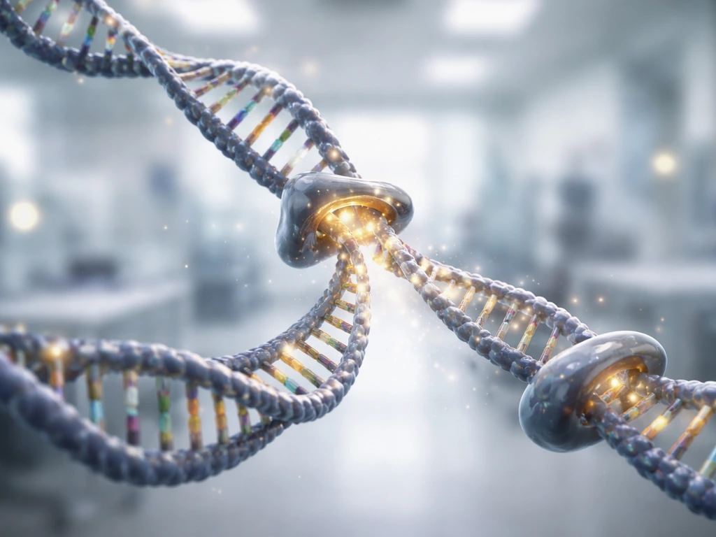
DNA is a double-helix molecule made of four chemical bases (adenine, thymine, guanine, cytosine) strung together in long sequences. In eukaryotic cells (like yours), most of that DNA is packed onto chromosomes inside the nucleus. DNA in cells is compartmentalized, and examples include the nucleus, which organizes DNA-containing biochemical reactions blank" rel="noopener noreferrer">packed onto chromosomes inside the nucleus. The nucleus helps the cell grow by housing the chromosomes the cell duplicates and uses when dividing. You also have a separate, smaller batch of circular DNA sitting inside your mitochondria, called mitochondrial DNA (mtDNA). Plants add a third location: chloroplasts. But in every case, we are talking about a chemical molecule, not a living structure that breathes, feeds, or expands on its own terms.
This matters because the word "grow" means something specific in biology. Growth refers to an increase in the size or number of biological structures, driven by cell division and followed by increases in cell size. DNA does not fit that definition. It is more like the instruction manual inside a factory than the factory itself. The manual does not grow; the factory uses the manual to build more factories.
DNA replication: the closest thing to DNA "growing"
If you want to find the moment when DNA quantity actually increases, look at S phase of the cell cycle. S stands for synthesis, and it is the specific window during interphase when the cell copies its entire genome. During interphase, a cell grows in preparation for division, and part of that process is copying the genome in S phase. Before S phase (during G1), the cell grows in size and stockpiles resources. During S phase, specialized enzymes unwind the double helix and build a complementary copy of each strand. By the end of S phase, the DNA content of that single cell has precisely doubled.
The machinery doing this work is impressively complex. An enzyme called helicase unzips the double helix at replication forks. Primase lays down short RNA primers, then DNA polymerases (including pol α, pol δ, and pol ε depending on the strand) add new nucleotides one at a time. DNA ligase seals gaps between the short Okazaki fragments made on the lagging strand, and topoisomerases prevent the DNA ahead of the fork from tangling up like a twisted garden hose. The error rate is around one mistake per one billion nucleotides copied, kept that low by proofreading activity built into the polymerases and a mismatch repair system that catches errors afterward.
Crucially, this entire process is tightly controlled. Cells only allow the genome to be copied once per cycle. After the G1-to-S transition, a set of molecular brakes (including CDK activity and a protein called geminin) blocks any replication origin from being licensed and fired again. This is the cell's way of making sure it makes exactly two copies, not three or four, before dividing.
Does DNA increase in your body over time?
Yes and no, depending on the scale you're looking at. Inside a single cell, DNA quantity stays constant unless that cell is preparing to divide. A cell sitting in G0 (the quiescent, non-dividing state) does not synthesize new DNA and its total DNA content doesn't change. In contrast, a cell actively cycling will double its DNA in S phase and then split it equally between two daughter cells at mitosis, so each daughter ends up with the same amount the mother cell started with.
At the tissue or organism level, the picture shifts. A growing child has more cells than a newborn, and more cells means more total DNA in the body. The cell wall (and the rest of the cell envelope) provides structure and a safe place to expand, so the cell can increase in size as it builds new material for growth. But that DNA increase comes entirely from cell division, not from any individual DNA molecule expanding. Meanwhile, in adult tissues, cell proliferation is carefully balanced with cell death so that total cell number stays roughly constant. Stem cells divide and generate new cells; old cells die off through apoptosis. The total DNA in your liver, for example, stays relatively stable because new hepatocytes replace old ones rather than piling up on top.
One nuance worth noting: mitochondrial DNA behaves a bit differently. Mitochondria can multiply by fission inside a cell independent of the nuclear cell cycle, so mtDNA copy number can shift somewhat independently of what the nucleus is doing. But even then, it is the cell controlling mitochondrial replication, not the DNA growing itself.
Growing DNA outside the body: PCR and cloning
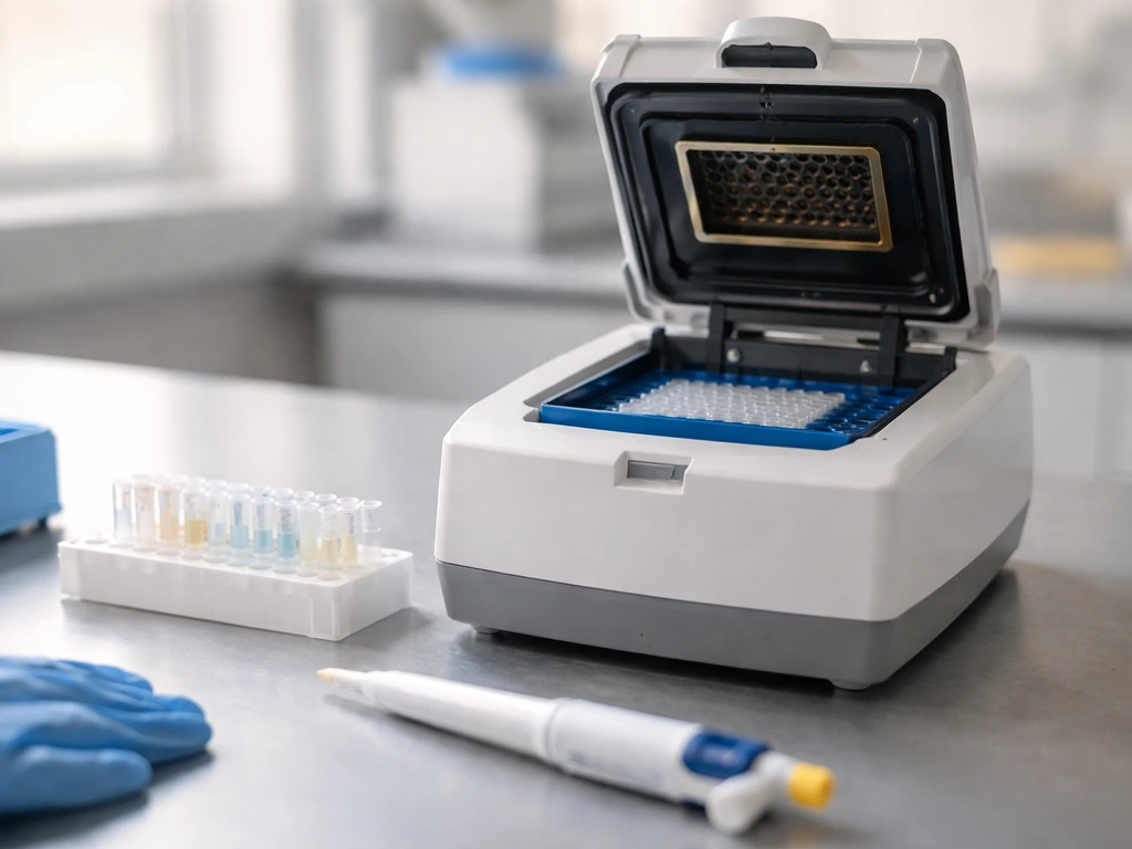
Scientists do routinely "grow" DNA in the lab, though they use words like amplify or propagate instead. The two main approaches are PCR (polymerase chain reaction) and molecular cloning, and they work on very different principles.
PCR: making millions of copies in hours
PCR takes a tiny DNA template and uses short sequences called primers, a heat-stable DNA polymerase (usually Taq polymerase), and a supply of free nucleotides to copy a specific target region over and over. Each cycle roughly doubles the number of copies, so after 30 cycles you can go from a handful of molecules to billions. Quantitative PCR (qPCR) tracks this amplification in real time using fluorescence: the cycle number at which the fluorescence crosses a threshold (called the Cq or Ct value) tells you how much starting DNA you had. The key point is that PCR still needs an enzyme and a template. The DNA is not growing; it is being copied by a molecular machine, just like inside a cell.
Molecular cloning: letting bacteria do the work
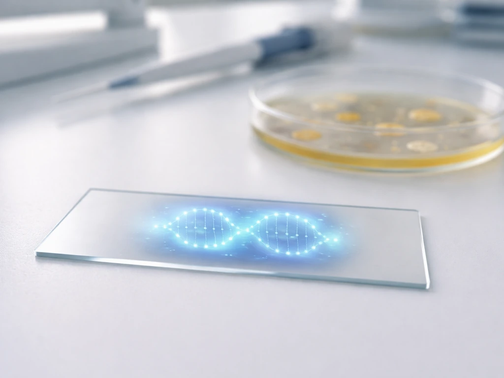
Molecular cloning takes a different approach. You insert your DNA fragment of interest into a vector, typically a small circular DNA molecule called a plasmid. The plasmid has its own origin of replication, a multiple cloning site where you slot in your fragment, and a selectable marker (like antibiotic resistance) so you can identify bacteria that successfully took up the plasmid. You then introduce the vector into bacterial host cells. Every time those bacteria divide, they replicate the plasmid along with their own chromosome, so your inserted DNA gets copied too. Grow a large culture of those bacteria and you end up with enormous quantities of your target DNA. Again: the DNA is not growing; living cells are copying it.
What stops DNA from replicating without limit?
Several overlapping constraints keep DNA replication from running out of control, both inside cells and in lab settings.
- Resource limits: DNA polymerases need a constant supply of deoxyribonucleotides (dNTPs, the building blocks). When dNTP pools run low, replication forks stall. Research shows that increasing dNTP levels can reduce fork stalling, which tells you that raw material availability is a genuine bottleneck.
- Enzyme availability: Helicase, primase, polymerases, ligase, and topoisomerases are all required in coordinated amounts. A shortage of any one of them slows the whole process.
- Licensing controls: Cells block re-replication within the same cycle using CDK signaling and geminin. Once an origin has fired, it cannot fire again until the next G1 phase.
- Replication stress checkpoints: When forks stall or DNA is damaged, the ATR-Chk1 signaling pathway kicks in. It suppresses new origin firing, stabilizes stalled forks, and can halt cell-cycle progression entirely until the problem is resolved.
- Cell senescence: Cells that accumulate too much damage may enter senescence, a permanent cell-cycle arrest typically at G1, where DNA replication simply does not start.
- In lab settings: PCR runs out of primers, nucleotides, or polymerase activity; bacteria in a cloning culture eventually hit nutrient and space limits, slowing division.
Common misconceptions worth clearing up
Misconception 1: DNA grows like an organism grows
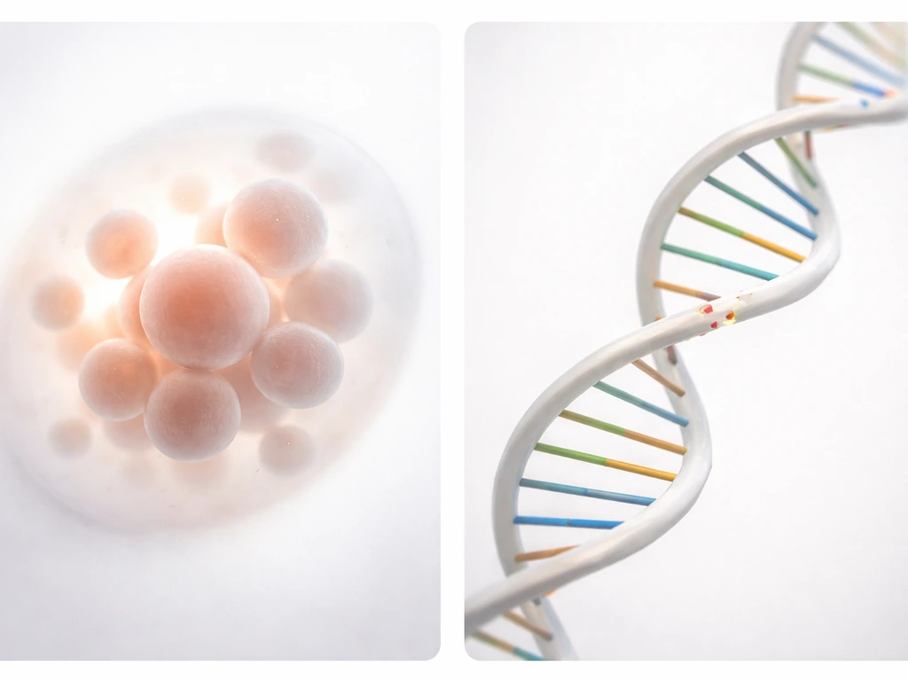
An organism grows because its cells divide and enlarge. DNA does not do either of those things independently. The sequence of a DNA molecule stays fixed unless a mutation occurs, and the physical length of a DNA strand does not increase simply because the organism gets bigger. What changes is how many copies of that DNA exist across all of the organism's cells.
Misconception 2: mutations mean DNA is "growing" or changing constantly
Mutations do occur during replication, but at a rate of roughly one error per billion nucleotides copied, thanks to polymerase proofreading and mismatch repair. Most lesions that block replication are not mutations; they get repaired before the fork moves on. DNA repair systems (base excision repair, nucleotide excision repair, mismatch repair, and others) constantly monitor and fix damage. So DNA is not randomly changing or expanding; it is being carefully maintained.
Misconception 3: DNA replicates itself
This one comes up a lot. DNA does not self-replicate. It cannot copy itself in a test tube sitting on a bench with nothing else around. Replication requires helicase, primase, polymerases, ligase, topoisomerases, a supply of dNTPs, ATP for energy, and cellular regulation that coordinates timing. DNA provides the template; the cell provides everything else. The two things are not the same.
Misconception 4: more DNA always means growth
A cell briefly has double the DNA it started with, right between the end of S phase and the completion of mitosis. This happens right between the end of S phase and the completion of mitosis. But that extra DNA is not "growth" in the biological sense; it is temporary duplication on the way to making two cells. After cytokinesis, each daughter cell is back to the starting DNA content. If you are curious about where exactly in the cycle this happens, that connects directly to the broader question of what phase the cell grows and whether all of that growth is DNA-related.
How to think about DNA changes over time (and how scientists measure them)
If you want to track DNA quantity in real life, here is what scientists actually do, at a conceptual level you can use to build a mental model.
| Method | What it measures | What it tells you |
|---|---|---|
| Flow cytometry with propidium iodide (PI) | Total DNA content per cell (fluorescence intensity) | Which cell-cycle phase each cell is in; 2C = G1, 4C = G2/M |
| BrdU or EdU pulse labeling | Newly synthesized DNA (analog incorporation during S phase) | How many cells are actively replicating DNA right now |
| qPCR | Amount of a specific DNA sequence in a sample | Whether a gene or sequence is present and how many copies |
| Replication timing sequencing | When specific genome regions are copied during S phase | Early vs. late replicating regions across the genome |
The flow cytometry approach is especially intuitive. Cells stained with propidium iodide glow brighter when they have more DNA. Plot the fluorescence of thousands of cells and you get two peaks: one at the 2C level (G1 cells with one diploid genome copy) and one at the 4C level (G2/M cells that have just finished replication). The trough between them represents cells currently in S phase, actively copying. This is the clearest visual proof that DNA quantity inside cells increases in a regulated, temporary, and purposeful way.
For a broader organism-level view, you can think about it this way: count all the cells in a tissue, multiply by the DNA content per cell, and you get total DNA in that tissue. During growth (say, a child developing), that number climbs as cell division outpaces cell death. In a stable adult tissue, it holds roughly steady. In aging or disease, things can get more complex, with some cells accumulating DNA damage, undergoing senescence, or even gaining extra chromosome copies (aneuploidy). But in healthy, typical tissue, the story stays clean: DNA increases only when cells replicate it, and it does so on a strict schedule.
Your practical takeaways
- DNA does not grow. It is a molecule that gets copied by cellular machinery under tightly regulated conditions.
- DNA quantity increases inside a cell during S phase of the cell cycle, when the genome is duplicated before cell division.
- At the organism level, total DNA increases as the number of cells increases during growth and development, not because individual molecules expand.
- In lab settings, DNA can be amplified via PCR (rapid, cell-free copying of a specific region) or propagated via molecular cloning (inserting DNA into a vector that replicates inside bacterial cells).
- Multiple systems, including checkpoint signaling, licensing controls, and resource limits, prevent DNA replication from running unchecked.
- Mutations happen but are rare and corrected by repair systems; they are not evidence of DNA "growing" or randomly changing.
- If you want to observe DNA quantity changes conceptually, think about the cell cycle: track a single cell from G1 through S phase into G2, watch the DNA content double, then follow it through mitosis as two daughter cells each receive half.
The big picture: asking whether DNA grows is actually a great way to sharpen your understanding of what growth really means in biology. Growth is not just "more stuff." It is a coordinated, resource-limited, cell-driven process. DNA is the blueprint that enables that process, not the thing doing the growing. But that same idea comes up in a more sci-fi way, with people asking can we grow dinosaurs from dna. Once that clicks, a lot of other biology, from how the cell cycle works to why organisms have size limits, starts to make a lot more sense.
FAQ
Does DNA “grow” during fertilization or early embryonic development?
Not by expanding itself. The embryo increases total DNA because cell divisions repeatedly enter S phase, doubling the genome each cycle so daughter cells inherit the same DNA amount as the parent cells at that stage.
If a cell grows larger in G1, does that mean its DNA grows too?
No. In typical cell cycles, G1 growth is mainly increases in cell size and biomolecule content, the genome is not copied yet. DNA quantity stays at the pre-replication level until S phase begins.
In cancer, does DNA start growing on its own?
Cancer is usually dysregulation of the cell cycle, not DNA self-growth. Replication can happen more often or in an abnormal pattern, leading to changes in total DNA per cell across time, including DNA damage and errors that can cause genome instability.
What is the difference between DNA amount (2C, 4C) and DNA length?
DNA length per genome does not increase just because the organism grows. The “2C to 4C” change reflects copying the same fixed-length genome, so each cell temporarily has more DNA molecules (twice the genome content) during and just after replication.
Does mitochondrial DNA increase the same way as nuclear DNA?
Not exactly. Mitochondria can change in number by fission and their genomes can be copied independently of the nuclear S phase, so mtDNA copy number can rise or fall without a direct one-to-one match with the cell cycle.
Can DNA replication happen without cell division?
Yes, DNA synthesis can occur while the cell fails to complete mitosis or cytokinesis, creating polyploid or multinucleated states. In that case, the DNA content per cell can increase temporarily, but it still requires replication machinery rather than “DNA growth.”
Why do DNA sequencing and PCR require DNA that already exists?
Because DNA cannot self-replicate. Sequencing and PCR need an input template, plus enzymes and energy. They measure or copy existing sequences, they do not create DNA from scratch.
Does PCR always double DNA every cycle?
Not perfectly. In ideal conditions it trends toward doubling, but reaction efficiency can drop due to primer limits, inhibitors, suboptimal annealing temperature, or template quality, which is why qPCR relies on calibrated interpretation rather than assuming exact 2x each time.
In molecular cloning, does the plasmid “grow” inside the tube?
The DNA amount increases because bacteria replicate the plasmid as they divide, but DNA alone in the tube will not multiply. Also, plasmid copy number per bacterium can vary by plasmid origin, changing total yield.
Can DNA damage make DNA look like it “grew” in experiments?
It can, indirectly. DNA fragmentation, replication stress, or abnormal cell cycle timing can alter how much DNA stains in assays like flow cytometry or how efficiently PCR works, so changes in measured signal do not always mean genuine successful replication of intact genomes.
How do researchers distinguish cells actively copying DNA from cells with more DNA total?
A common approach is to use DNA content staining plus cell cycle gating. For example, cells in S phase often occupy intermediate DNA-content levels between the pre- and post-replication peaks, while cells in G1 and G2/M cluster at distinct higher and lower fluorescence intensities.
Next Article
During Which Period Is DNA Replicated and Cells Grow
DNA replicates in S phase, while cells grow mainly in G1 and prepare in G2 before mitosis.