You can grow skin cells from stem cells by guiding pluripotent stem cells through a staged chemical signaling process that mimics what happens in a developing embryo. The key steps are: start with the right stem cell source, expose those cells to retinoic acid and BMP4 to push them toward an epidermal (skin) fate, culture them on collagen-coated surfaces to enrich for keratinocytes, then verify your results with flow cytometry and marker staining. Done properly, published protocols reach 80–95% KRT14-positive keratinocyte-like cells within 28 days. Here is how that whole workflow fits together, and what to watch for at each stage.
How to Grow Skin Cells from Stem Cells: A Practical Guide

Marcus Whitmore
1 Jun 2026
What you are actually trying to do here
"Growing skin cells from stem cells" involves two distinct biological processes running in parallel: cell proliferation (cells dividing and making more of themselves) and cell differentiation (cells committing to a specific identity, in this case an epidermal keratinocyte). You need both, but in the right sequence. Think of it like baking bread: the yeast has to first activate before it can rise. You cannot skip directly to the rise.
The target cell type here is the keratinocyte, the dominant cell of the skin's outer layer (the epidermis). Keratinocytes express characteristic structural proteins like KRT14, KRT5, and eventually involucrin and filaggrin as they mature. Your job in the lab is to convince a pluripotent stem cell, which could theoretically become any cell in the body, to travel down the specific developmental path that embryonic skin cells follow. That path goes: pluripotent stem cell → ectoderm → surface ectoderm/epidermal progenitor → keratinocyte progenitor → mature keratinocyte. Each arrow in that sequence requires the right molecular signals at the right time, much like a train switching tracks at each junction.
This is also a useful case study for understanding how growth is constrained in biology more broadly. Cells do not grow indefinitely. At each differentiation stage, growth signals and physical environment literally reshape what is possible. The culture dish, the substrate, the oxygen concentration, and the cocktail of growth factors all act as physical and chemical walls that channel cells toward (or away from) your intended outcome. That tension between growth potential and environmental constraint is exactly what makes this process so interesting to study.
Choosing your starting stem cell source
Your stem cell source shapes everything downstream: cost, ethical considerations, ease of reprogramming, and the quality of the cells you ultimately produce. There are three main options you will encounter in the literature.
| Stem Cell Type | How You Get Them | Main Advantage | Main Limitation |
|---|---|---|---|
| Human induced pluripotent stem cells (hiPSCs) | Reprogrammed from adult somatic cells (e.g., skin fibroblasts or blood cells) | Patient-specific; avoids embryo use; broadly used in published keratinocyte protocols | Reprogramming adds time and cost; quality varies between lines |
| Human embryonic stem cells (hESCs) | Derived from donated embryos | Well-characterized; many protocols originally validated on these | Ethically regulated; requires specific oversight and approvals |
| Adult epidermal stem cells | Biopsied directly from skin tissue | Already committed to skin fate; shorter differentiation needed | Limited availability; harder to expand; not pluripotent |
For most educational and research protocols today, hiPSCs are the default. They are the subject of most published keratinocyte differentiation papers, and they sidestep many of the ethical restrictions associated with hESC use. If you are learning the workflow conceptually, though, know that the signaling biology is virtually identical between hiPSC- and hESC-based protocols. One note: working with any human stem cell line, including hiPSCs, typically requires institutional review board (IRB) oversight, and sometimes additional Institutional Biosafety Committee (IBC) approval. FDA warning communications also note that an IND must be in effect to lawfully distribute stem-cell products for clinical use and highlight enforcement around unapproved stem-cell products. Do not skip that step.
The differentiation steering wheel: signals that drive skin fate
Signaling molecules are the actual "steering wheel" here. To move a pluripotent cell toward keratinocyte fate, you have to activate some pathways and block others, in a timed sequence. Here is the core logic of what published protocols do.
Stage 1: Push toward ectoderm with retinoic acid and BMP4
The first major move is exposing your pluripotent stem cells to retinoic acid (RA) and BMP4. Retinoic acid promotes ectodermal (surface layer) fate, while BMP4 suppresses the alternative neural pathway that ectodermal cells might otherwise take. Without BMP4, you risk generating neurons instead of skin cells. Together, they drive cells toward surface ectoderm, the embryonic tissue that gives rise to skin. You can think of RA as pointing the car toward ectoderm and BMP4 as blocking the neural off-ramp.
By around day 14 of this induction, you should see clear molecular evidence of commitment: pluripotency markers OCT4 and NANOG should drop significantly, while keratinocyte progenitor markers KRT18 and p63 should rise. A canonical pathway in the literature uses retinoic acid and BMP4 to promote ectoderm fate from hPSCs while blocking neural fate, generating keratinocyte progenitor-like cells assessed by marker changes around day 14 Directed differentiation of human pluripotent stem cells into epidermal keratinocyte. This is your checkpoint that the RA+BMP4 treatment actually worked. If those markers are not shifting, something went wrong in the induction, and it is worth re-checking your reagent concentrations and timing before moving on.
Stage 2: Fine-tune with TGF-β inhibition, WNT modulation, and Notch inhibition

More sophisticated staged protocols add extra layers of control. BMP4 used alongside a TGF-β inhibitor (such as SB431542) early in differentiation pushes the epidermal outcome further. WNT pathway activation, for example using the small molecule CHIR99021, is required at the epidermal fate stage but needs to be carefully timed: the wrong amount of WNT signaling at the wrong time sends cells off-course. Later in the protocol, Notch pathway inhibition (using a compound like DAPT) helps drive cells into later keratinocyte stages. Each of these is essentially a molecular gate that cells must pass through in the right order, like checkpoints on a train line.
A critical timing nuance worth knowing: TP63, a transcription factor that marks epidermal progenitors, can be highly expressed by day 8, while KRT14, the classic keratinocyte marker, may not appear until around day 11 or later. If you check for KRT14 at day 8 and see low signal, that is not necessarily failure. It just means you are still in the progenitor phase. Staged marker tracking is much more informative than a single snapshot.
Culture conditions: medium, oxygen, and timing
Getting the chemical signals right is only part of the picture. The physical culture environment matters enormously, and several published failures trace back to getting these conditions wrong rather than the signaling protocol itself.
- Oxygen tension: Maintain iPSCs under low-oxygen (hypoxic) conditions before differentiation, then switch to atmospheric oxygen (~20%) during the keratinocyte differentiation protocol itself. This switch is not optional in some published protocols; it affects cell fate outcomes.
- Medium composition: Keratinocyte growth medium (KGM) formulations are commonly used during the expansion and maturation phases. Note that keratinocytes propagated in serum-free, feeder-free conditions can fail to form stratified, multilayered epidermis in 3D models, so medium composition directly affects how far the cells can mature.
- Calcium concentration: While calcium is important for keratinocyte stratification, it is not the sole gate for early ectodermal commitment. Some defined protocols tolerate standard calcium levels during early induction stages without disrupting fate decisions.
- Timing: Marker progression follows a predictable schedule in published protocols. KRT14+ cells reported at roughly 67% by day 7, 73% by day 14, 88% by day 21, and 95% by day 28 in defined-condition protocols. Track at multiple timepoints rather than relying on endpoint-only readouts.
Mimicking skin with scaffolds, substrates, and 3D culture
Real skin is not a flat plastic dish. Keratinocytes in the body sit on a basement membrane made of proteins like collagen and laminin, and they feel mechanical forces that affect how they behave. Getting close to those physical conditions in vitro is what separates a basic 2D culture of keratinocyte-like cells from something that actually resembles functional skin.
2D culture on collagen-coated surfaces
The simplest and most widely used approach is growing cells on dishes coated with collagen type I and collagen type IV. These extracellular matrix (ECM) proteins mimic the basement membrane and, importantly, KRT14-positive keratinocyte progenitors adhere to them preferentially and rapidly. This gives you an adhesion-based enrichment step: committed keratinocyte-like cells stick, while off-target or less committed cells do not. Published protocols report enrichment up to 80–90% KRT14+ using this approach. You can also achieve rapid primary keratinocyte expansion on hydrogel substrates tuned to match the elasticity of physiological skin niches, which promotes proliferation and functional behavior over stiffer or more artificial surfaces.
Substrate stiffness and laminin
Mechanical cues are real biological inputs, not just technical details. Substrate stiffness affects keratinocyte colony formation and stratification readiness. Softer substrates tuned to match the mechanical properties of real skin promote proliferation and self-renewal better than rigid plastic. Laminin-332, another key basement membrane protein, strongly affects how keratinocytes adhere and migrate: polymerized laminin-332 supports tight adhesion while suppressing excessive migration, which matters when you want cells to settle and organize rather than scatter.
3D air-liquid interface (ALI) culture
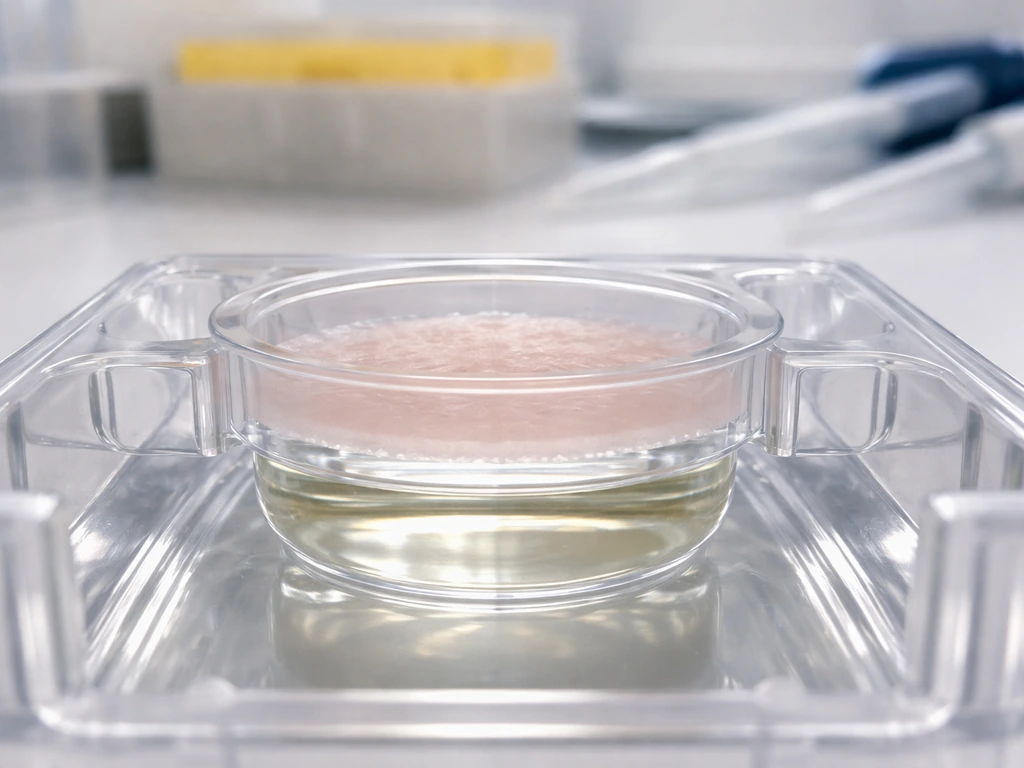
If you want keratinocytes to form actual epidermal strata, not just grow as a flat monolayer, you need to move into 3D air-liquid interface (ALI) culture. In an ALI model, keratinocytes are seeded onto a permeable membrane or scaffold, then the apical surface is exposed to air while the basal side remains in contact with liquid medium.
Published studies using hESC- and iPSC-derived keratinocytes in ALI culture with sequential high-to-low humidity transitions have produced epidermal strata with barrier-like properties that closely resemble normal skin. This is where the cells actually stratify: basal progenitor layer at the bottom, progressively more differentiated layers above, eventually cornified (dead, protective) cells at the top.
If stratification fails in your model, check whether your keratinocytes were propagated in serum-free feeder-free conditions, since that combination has been documented as a cause of stratification failure in reconstituted skin models.
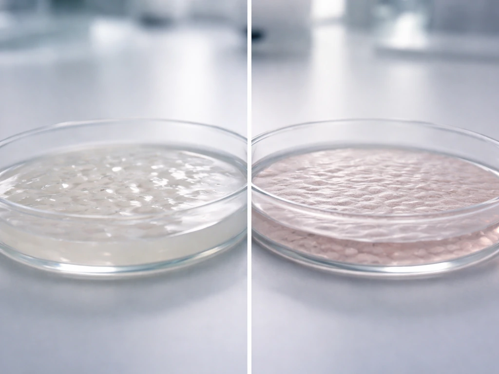
How to confirm you actually made skin cells
This step is non-negotiable. Morphology alone (cells looking skin-like under a microscope) is not sufficient to confirm identity. You need molecular evidence. Here is the core readout toolkit.
Markers to track at each stage
| Stage | Markers Expected ON | Markers Expected OFF |
|---|---|---|
| Pluripotent stem cells (start) | OCT4, NANOG, SOX2 | KRT14, KRT5, p63, KRT18 |
| Ectodermal/progenitor commitment (~day 14) | KRT18, p63, TP63 | OCT4, NANOG |
| Early keratinocyte (~day 8–11) | TP63, early KRT14 | Neural markers (PAX6) |
| Maturing keratinocyte (~day 21–28) | KRT14, KRT5, KRT15, KRT10, involucrin (IVL) | OCT4, NANOG, SOX2 |
| Terminally differentiated keratinocyte | Involucrin, filaggrin, KRT10 | Basal markers at high level |
Assays to use
- Flow cytometry: The most quantitative readout. Measure KRT14, KRT10, and involucrin (IVL) positivity at days 7, 14, 21, and 28. Published defined-condition protocols report KRT14+ rising from ~67% at day 7 to ~95% at day 28. Use this to confirm your protocol is on track, not just at the endpoint.
- qRT-PCR: Measure gene expression for KRT5, KRT8, KRT14, KRT15, involucrin, and filaggrin. Also confirm pluripotency marker loss (NANOG, OCT3/4, SOX2 should be absent). This gives you transcript-level evidence of differentiation.
- Immunofluorescence staining: Visualize protein distribution in cells directly. Look for KRT14 and p63 co-expression in the same cells (TP63/KRT14 double-positive is a key acceptance criterion in published quality control steps). Morphology combined with marker localization is far more informative than either alone.
- Acceptance criterion: A commonly used quality threshold is the fraction of TP63 and KRT14 double-positive cells. If your culture does not meet this threshold (typically target >80% KRT14+ after collagen enrichment), do not proceed to downstream experiments.
Common failures and how to troubleshoot them
Even well-designed protocols fail. Here are the most common problems, what causes them, and what to do.
- Expecting KRT14 too early: p63 can be positive by day 10 at around 30% of cells, while KRT14 is still low at that stage. This is normal progression, not failure. Do not abandon the protocol at day 10 because KRT14 looks low. Check TP63 first; if it is rising, you are on track.
- Mixed or off-target cell populations: If your differentiation produces a mixture of cell types (some neural, some mesenchymal, some still pluripotent), it typically means BMP4 concentration was insufficient to block neural fate, or WNT timing was off. Re-check your reagent concentrations and switch timing. Also review whether you removed undesired populations during differentiation; this cleanup step is documented as essential in published protocols.
- Pluripotency markers that won't go away: If OCT4 and NANOG are still strongly expressed past day 14, your induction did not work. Possible causes: RA or BMP4 was degraded or under-dosed, or the iPSC culture had quality issues. Verify reagent freshness and iPSC pluripotency quality before restarting.
- Poor stratification in 3D ALI models: If your cells don't form multilayered strata at the air-liquid interface, check whether they were propagated in serum-free feeder-free conditions. This specific combination has been shown to block stratification in reconstituted skin models. Consider adjusting medium composition or adding feeder support.
- Low KRT14+ yield after collagen enrichment: If you're below 80% KRT14+ after the collagen I/IV adherence step, either your differentiation was incomplete, or the enrichment step timing was off. Confirm that the differentiation phase reached the right progenitor stage before attempting adhesion-based selection.
- Ignoring regulatory requirements: If you are planning to work with human stem cells in a formal research context, not just reading about it, IRB review may be required for human subject research, and IBC approval is often needed for work with human-derived cells. Clinical use of any stem-cell-derived product without FDA oversight (including an active IND) is illegal. Start with institutional compliance before starting the biology.
Realistic next steps for learning this workflow
If you are a student or curious learner trying to understand this topic rather than run a lab experiment, the clearest path forward is to work through published open-access protocols. It is the same idea, just with different targets, to ask whether stem cells can be guided to form kidney tissue can you grow a kidney from stem cells. Look for papers describing hiPSC-to-keratinocyte differentiation using defined conditions; these typically include full materials lists, day-by-day timelines, and expected marker data at each checkpoint. Reading those alongside this overview will make the biology concrete. Pay attention to the control experiments: how authors confirm that their differentiation actually worked, and what they do when early markers look ambiguous.
If you are an advanced reader aiming to run a version of this yourself in a licensed research setting, the practical starting point is validating your hiPSC line quality first. A poorly maintained or low-quality iPSC line will fail at the induction stage no matter how good your signaling cocktail is. Once you have a clean, pluripotency-confirmed starting population, follow a staged protocol with checkpoint flow cytometry at days 7, 14, and 21 before committing to the full 28-day run. The collagen I/IV enrichment step is your single best tool for cleaning up mixed populations before verification.
It is also worth zooming out and appreciating where skin sits among the organs that researchers are working to build from stem cells. Skin is considered one of the more tractable targets because keratinocytes are relatively robust, have well-established culture methods, and don't require the same vascularization complexity as organs like kidneys, hearts, livers, or pancreases.
Those principles also inform how researchers ask, can you grow a heart from stem cells, using staged signaling to drive cardiac identity hearts. Kidney tissue engineering is a much harder goal, but the same stem-cell control logic and checkpoint-based verification apply organs like kidneys. If you are wondering whether stem-cell approaches can extend beyond skin to something like liver tissue, you can ask a similar question: what exact lineage signals and culture constraints are required for hepatocyte fate livers.
That relative accessibility makes skin an excellent entry point for understanding stem cell differentiation biology more broadly, and the same principles of staged signaling, environmental constraint, and marker-based verification apply to those more complex organ systems too.
The core lesson from all of this is that "growing" skin cells is really a story about control: controlling which signaling pathways are active, controlling the physical environment, controlling the timing of each stage, and then measuring carefully to confirm the cells are where you think they are. Researchers can use the same staged differentiation and checkpoint mindset to ask whether you can grow a liver from stem cells can you grow a liver from stem cells. That is the same fundamental logic that governs how any complex living structure grows, from a crystal face to an embryonic organ. The signals set the direction; the environment sets the constraints; growth follows within those boundaries.
FAQ
Do I have to use retinoic acid and BMP4, or can I switch to other signals to reach keratinocytes?
You can sometimes substitute related pathway modulators, but RA (to support ectoderm) and BMP4 (to suppress the neural off-ramp) are central in most successful keratinocyte-induction schemes. If you change them, you should add an extra validation step earlier (day 7 to 10) to confirm ectoderm commitment markers are shifting, not just later KRT14 signals.
If OCT4 and NANOG drop but KRT18 or p63 do not rise by day 14, what is the most likely issue?
The most common causes are mistimed dosing or cell-health problems from the start (over-confluence, stressed colonies, or uneven differentiation). Re-check starting colony density and viability, then verify that the induction medium was refreshed on schedule and that the actual incubation conditions (temperature, oxygen tension, and handling times) matched the protocol.
Why do my cells express TP63 early but show weak KRT14 later?
This pattern often means you are in the epidermal progenitor window rather than fully transitioning to keratinocyte maturation. The article’s timing nuance matters, TP63 can peak around day 8 while KRT14 may not appear until day 11 or later. Use a time-course readout (for example days 8, 11, 14) rather than relying on a single early checkpoint.
Should I optimize using a 2D culture only, or can I go straight to 3D air-liquid interface (ALI)?
In practice, ALI usually works best after you have already enriched keratinocyte-like progenitors. If you seed ALI too early with mixed populations, stratification often fails or becomes patchy. A practical approach is to do the collagen I/IV enrichment and marker confirmation first, then transfer to ALI.
What are the most informative markers to confirm identity beyond KRT14 positivity?
KRT14 is necessary but not sufficient. Pair it with progenitor and maturation markers such as KRT5, involucrin, and filaggrin, depending on your stage, and confirm barrier-associated behavior in ALI when relevant. Also include a negative control panel (markers of neural or mesenchymal fates) to detect cross-lineage drift.
How do I know whether poor stratification is due to culture conditions versus differentiation signaling?
Use the checkpoints to localize the failure. If day 14 or day 21 shows expected epidermal/progenitor marker shifts, but ALI does not stratify, the issue is more likely physical culture variables such as serum-free, feeder-free propagation, membrane seeding density, or ALI humidity and transition timing. If even early markers fail to shift, signaling is the more likely culprit.
Is collagen I/IV coating the only ECM option, and how do I decide on ECM concentration?
It is not the only option, but collagen I/IV is widely used because it provides adhesion-based enrichment of KRT14-positive progenitors. ECM concentration and coating uniformity can strongly affect attachment and later differentiation, so treat it as a tunable parameter and keep it consistent within an experiment. When testing alternatives, first confirm adhesion kinetics and early marker enrichment before moving to ALI.
What is a common mistake when using flow cytometry checkpoints in this workflow?
A frequent problem is gating based only on healthy-appearing cells without confirming single-cell gating and viability. Keratinocyte-like differentiation cultures can contain clumps and dead cells that distort quantification. Always include viability dye and singlet gating, and run a consistent unstained and isotype or FMO control strategy.
Can I start from any hiPSC line, or is line quality a major bottleneck?
Line quality is a major bottleneck. Even with correct signaling, poorly maintained or partially differentiated pluripotent lines can derail commitment. A practical decision aid is to confirm pluripotency and differentiation competence before running the full induction, then compare your day 7 checkpoint outcomes across lines to decide whether the bottleneck is upstream.
Do oxygen levels and incubation conditions matter, or is it mostly the chemical cocktail?
Incubation conditions matter, especially oxygen tension and handling time, because they influence differentiation kinetics and stress responses. If you see slow progression or inconsistent checkpoint shifts, correct environmental variables first (temperature control, CO2 settings, equilibration times), then revisit dosing timing and medium change frequency.
What should I do if early markers look “ambiguous,” but later KRT14 eventually appears?
Ambiguity early is not always failure, but you should decide based on trend and downstream consistency. If OCT4/NANOG decreases and epidermal progenitor markers rise by days 14 to 21, then proceed. If KRT14 shows up late but stratification in ALI remains poor, focus on enrichment quality and the physical culture transitions rather than repeating only the signaling stage.
Are these methods safe to attempt outside of a licensed research setting?
Work with human stem cells typically requires institutional oversight and compliance with biosafety and ethics rules (for example IRB and sometimes IBC approval). Beyond regulations, practical safety depends on your facility’s capabilities for cell handling and waste disposal, so do not attempt the workflow without appropriate authorization and training.
Next Article
Can You Grow a Liver From Stem Cells? What’s Possible
Learn what growing liver tissue from stem cells can mean, what works now, and why real transplant livers remain hard.