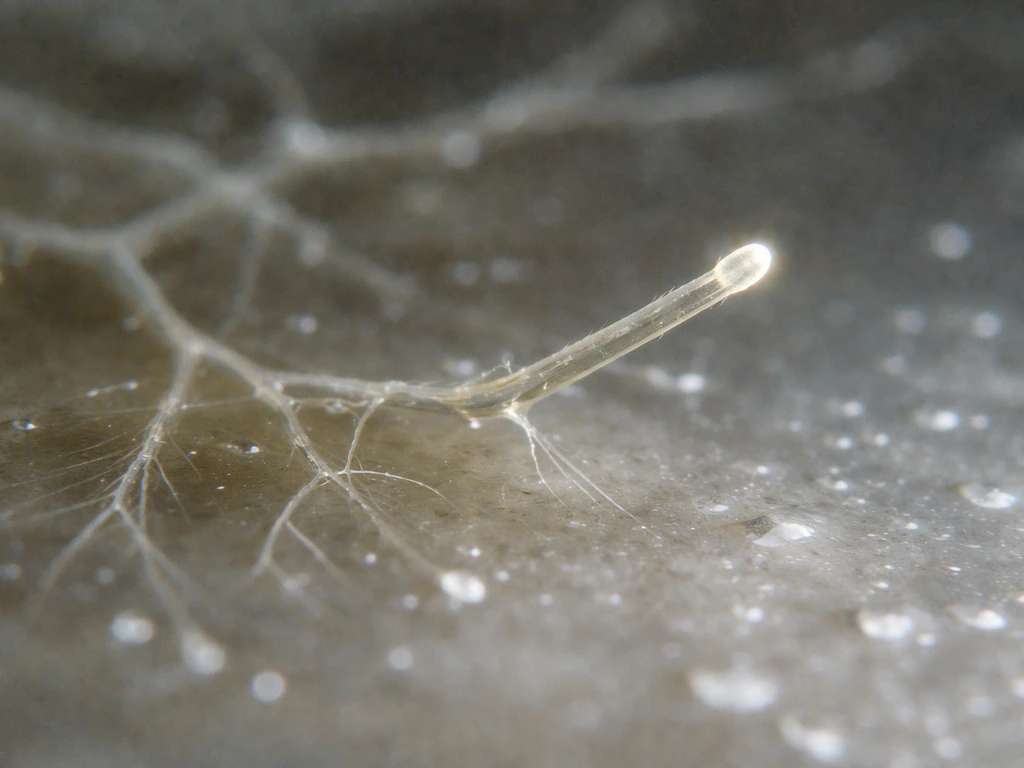
Hyphae grow exclusively at their tips. The rest of the tube stays locked in place while a tiny zone at the very front, just a few micrometers wide, pushes outward, lays down new cell wall, and drags the whole filament forward. That one fact, tip-only growth (called apical growth), explains almost everything else about how fungi colonize soil, food, wood, and living tissue so effectively.
How Do Hyphae Grow: Step-by-Step Biology and Conditions

Marcus Whitmore
16 May 2026
What hyphae are and what 'growth' actually means here
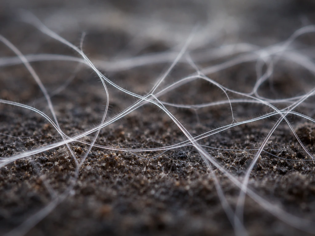
A hypha (plural: hyphae) is the basic structural unit of a filamentous fungus. Think of it as a long, branching tube, usually between 2 and 30 micrometers in diameter, that extends through a substrate and collectively forms mycelium, the white fuzzy network you see on bread mold or rotting wood. Each hypha is surrounded by a rigid cell wall made largely of chitin and glucans, with a plasma membrane inside, and cytoplasm streaming through the middle.
Fungal growth is not the same as what happens when an animal cell divides or when a plant root elongates. Hyphae do not lengthen by adding new cells along their sides. Instead, the whole extension event is polarized: the fungus concentrates its building machinery at one end and pushes that end forward continuously. A fast-growing hypha like Neurospora crassa can extend at rates of roughly 24 micrometers per minute under ideal conditions. That is surprisingly quick for something operating at a microscopic scale.
Understanding this is also key to appreciating how mycelium grows as a network, a topic worth exploring on its own. You can think of this network growth as the coordinated extension and branching of many hyphae working together in the same substrate how mycelium grows as a network. But here we are zooming all the way in to one individual hypha and asking: what is actually happening at the molecular level that makes the tip move?
Apical growth: how the tip actually pushes forward
Picture the hyphal tip as a tiny construction zone. The cell wall there is thin and plastic, not yet fully cross-linked, which allows it to yield to outward pressure. That pressure is turgor, the hydrostatic pressure inside the hypha generated by the accumulation of solutes (ions, sugars, amino acids) that draw water into the cell by osmosis. Turgor is the main physical force driving the tip outward, essentially inflating the soft wall material like the end of a balloon.
Turgor alone would just pop the cell if new wall material were not being deposited simultaneously. The trick is that wall-building enzymes and the vesicles carrying wall precursors are all converging on the tip at the same moment turgor is pushing out. The tip stays soft enough to expand but gets reinforced almost instantly afterward. When osmotic stress hits, this balance breaks down fast: experiments in Aspergillus nidulans show that salt stress can cut hyphal extension rates by around 60% and cause the apical growth zone to lose its organized structure entirely.
At the center of all this activity sits a structure called the Spitzenkörper (SPK), which translates loosely as 'tip body.' It is a phase-dark cluster of vesicles and polarity proteins visible under a standard light microscope right at the apex of growing hyphae in both Ascomycetes and Basidiomycetes. The SPK acts like a distribution hub: vesicles loaded with enzymes, membrane, and wall-building cargo arrive here, queue up, and then fuse with the plasma membrane at the very tip. Where the SPK is, growth is happening. If the SPK shifts or disappears, the growth direction changes or stops.
Getting materials to the tip: transport, streaming, and trafficking
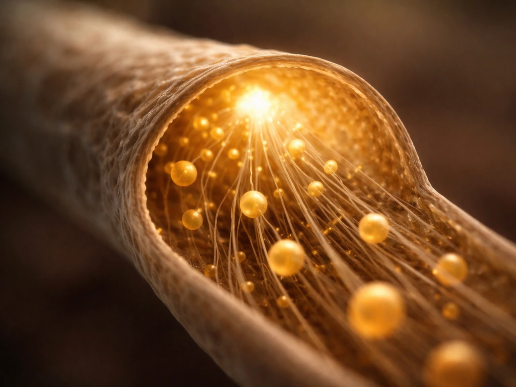
Moving vesicles from where they are made (in the endoplasmic reticulum and Golgi near the nucleus, which can be hundreds of micrometers behind the tip) to the apex requires a dedicated logistics system. Fungi use a combination of microtubule-based long-distance transport and actin-based short-range delivery, and the two systems hand cargo off to each other like a relay.
On the microtubule tracks, motor proteins do the hauling. Kinesin-1 moves secretory vesicles toward the tip at speeds of roughly 7 to 10 micrometers per second. Kinesin-3 handles early endosomes at somewhat slower speeds of about 2 to 7 micrometers per second. Dynein runs cargo in the opposite direction, away from the tip, which is important for recycling and signaling. In the corn smut fungus Ustilago maydis, a coordinated three-motor system involving Kin3, dynein, and conventional kinesin keeps this bidirectional traffic organized on polarized microtubule arrays.
Once vesicles get close to the tip, they transfer to actin cables for the final stretch. Myosin V motors (in Aspergillus, this is MyoE) take over and walk the vesicles to the apical cortex. When MyoE is genetically disabled, RAB11-positive secretory vesicles scatter away from the tip and the SPK loses its coherence. That tells us actin-myosin V handoff is not optional; it is the last critical step before exocytosis deposits cargo into the growing membrane.
The Spitzenkörper itself concentrates several key trafficking proteins, including Rab11/Ypt-31 and Sec4, both markers of late secretory vesicles. Superresolution live imaging has confirmed that these vesicles are not just drifting; they arrive at the SPK in distinct fast pulses, accumulate briefly, and then are released to fuse with the plasma membrane. The whole system looks remarkably like a just-in-time delivery operation, and if any link in the chain breaks, growth stalls.
Building the cell wall at the growing front
The fungal cell wall is mostly chitin (a tough polymer also found in insect exoskeletons) woven together with beta-glucans and other polysaccharides. At the tip, none of this is fully assembled yet; the wall is thin, extensible, and constantly being added to. The enzymes that do the building, chitin synthases, are delivered to the tip inside small membrane vesicles called chitosomes. This process is controlled in part by membrane-associated chitin synthase, and chitin synthase in a zymogen form has been detected in microvesicles called chitosomes that transport the enzyme to the hyphal tip. These microvesicles carry the enzymes in a dormant (zymogen) form and only activate once they fuse with the membrane at the right location.
Different classes of chitin synthase go to different places. In Aspergillus nidulans, ChsB (class III) and CsmA (class V) both depend on conventional kinesin for microtubule-based transport to the hyphal tip and to septation sites. Without that transport, chitin deposition becomes mislocalized and the tip loses structural integrity. Think of it as the difference between pouring concrete in the right spot versus slopping it randomly.
As vesicles unload their cargo at the tip and the wall starts to form, the newly deposited chitin gets cross-linked and stiffened by glucan synthases and other remodeling enzymes. This progressive hardening moves the 'soft zone' constantly forward. The subapical region, just a few micrometers behind the tip, is where vesicle contents begin to be released and wall material starts consolidating. Live-imaging studies tracking GFP-tagged chitin synthase vesicles have observed them advancing toward the tip and then beginning to break apart in the subapical zone, exactly where cargo release would be expected.
Direction, branching, and the internal organization that keeps growth on track
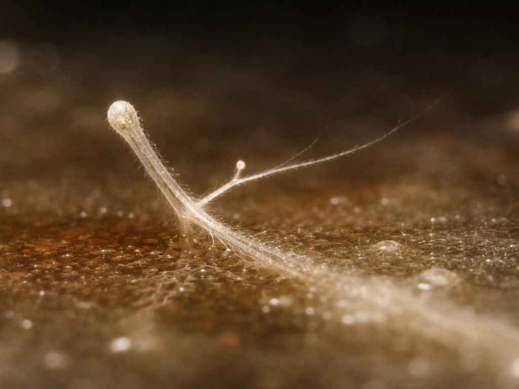
How does a hypha know which way to grow? Once you know what hyphae are signaling and building at the tip, the same principles help explain how do they grow in practice dates how do they grow. That same tip growth logic also explains how hyphae extend and expand as they grow through new territory How does a hypha know which way to grow. The short answer is polarity maintenance: a set of signaling proteins (Rho GTPases, polarisome components, and formin proteins) mark the tip as the active growth site and recruit everything else there. The Spitzenkörper position is itself a readout of this polarity axis. When the SPK is centered at the very tip, the hypha grows straight. This same apical growth program is what ultimately determines how a fungus can grow so effectively across different substrates. When it drifts to one side, the hypha curves. When it disappears, growth stops.
Branching is how fungi dramatically expand their reach without each individual hypha having to travel everywhere. A branch usually initiates just behind a septum or at a point of mechanical or chemical signaling. The cytoskeleton reorganizes to establish a second polarity axis, a new SPK forms, and a fresh growth zone opens. Under resource deficiency in Neurospora crassa, branch diameter shrinks from the normal 10 to 20 micrometers down to around 6.5 micrometers and branch growth rates fall from 24 to about 6.7 micrometers per minute, showing that branching is tightly coupled to available resources.
Septa, the cross-walls that divide a hypha into compartments, are a separate process from extension. Septum formation involves four well-defined steps: selection of the septation site, assembly of a contractile actomyosin ring, inward plasma membrane invagination, and deposition of new wall material (including chitin) across the ring. Septins (ring-forming GTP-binding proteins) mark the septation site and help coordinate the whole process. Once a septum matures, Woronin bodies can plug the pore in it, which is how a hypha seals off damaged compartments without losing cytoplasm from the whole network. It is a simple but elegant damage-control mechanism.
What speeds up or shuts down hyphal growth
Hyphal growth is highly sensitive to environmental conditions. Getting any one of these factors wrong can slow growth to a crawl or stop it entirely.
| Factor | Effect on growth | Practical notes |
|---|---|---|
| Water / humidity | Essential for turgor; drought stops growth immediately | Hyphae in dry substrates retract or go dormant |
| Temperature | Most species have a narrow optimal range (20-30°C for many saprophytes) | Too cold slows enzyme kinetics; too hot denatures proteins |
| Nutrient availability (C and N sources) | Carbon fuels energy and wall synthesis; nitrogen drives protein production | Deficiency reduces branch rate and diameter significantly |
| pH | Most fungi prefer mildly acidic conditions (pH 4-7) | Extreme pH disrupts wall enzymes and ion gradients |
| Oxygen / CO₂ | Most filamentous fungi are aerobic; low O₂ limits ATP and growth | High CO₂ can redirect or inhibit growth depending on species |
| Osmotic stress | High solute concentrations reduce turgor and disrupt tip organization | Salt stress cuts extension rates by ~60% in Aspergillus nidulans |
| Substrate / surface mechanics | Physical substrate guides direction and can resist or support expansion | Hard, dry, or toxic surfaces impede hyphal penetration |
Osmotic stress is worth calling out specifically because it hits two things at once: it reduces turgor (so the physical force driving tip expansion drops) and it triggers an osmotic MAPK signaling cascade that redirects cellular energy toward solute accumulation and ion uptake rather than wall synthesis. The cell is essentially choosing survival over growth, which makes sense but means growth can stay suppressed until conditions improve.
How to observe and troubleshoot hyphal growth in practice
If you want to actually watch hyphae grow, a compound microscope at 40x or 100x with a simple slide culture is enough to see the basics. Grow your fungus on a thin agar slab cut to slide size, cover it with a coverslip, and place it in a humid chamber. You can often watch tip extension happening in real time, especially in fast-growing species like Neurospora or Aspergillus. What you are looking for:
- A clear, smooth, dome-shaped apex with no visible granules right at the very tip (the granule-free zone marks the SPK region in many species)
- The rate of tip advance, which you can measure by marking position at timed intervals using a stage micrometer or calibrated image software
- Branching events, which typically appear as small bulges just behind septa before a new tip pushes out
- Septum formation, visible as faint transverse lines forming behind the advancing tip as the hypha ages
- Any change in direction, curving, or hesitation in the tip, which often signals a local environmental cue or physical obstacle
For more detailed work, the lipophilic dye FM4-64 (and related dyes like FM1-43) stains vesicle membranes with low toxicity and lets you track the apical vesicle cluster, including SPK dynamics, in living hyphae using confocal microscopy. It has been validated across a wide range of fungal species and is one of the most practical tools available for visualizing the vesicle traffic described above.
Troubleshooting common growth problems
If hyphal growth is sluggish or stalled, work through the environment before assuming a genetic or contamination issue. The most common causes and fixes:
- Check moisture first. If agar or substrate is drying out, hyphae will stop. Keep humidity high and seal plates with parafilm or tape.
- Verify temperature. A few degrees outside the optimal range can halve extension rates. Use a calibrated thermometer near the culture, not just the incubator display.
- Assess nutrient medium. If you see very thin, stunted branches, you may have nitrogen or carbon limitation. Compare to a rich-medium control.
- Check pH. If using liquid media, pH can drift during growth as the fungus consumes nutrients or excretes acids. Buffer the medium or test it with strips.
- Ensure adequate gas exchange. Tightly sealed containers with no air exchange will deplete oxygen. Use vented lids or breathable film.
- Look for osmotic stress. If you have added salts or other solutes, these may be suppressing turgor. Diluting the medium or switching substrates can restore growth.
- Examine tip morphology. If tips look swollen, irregular, or are branching excessively without extending, this often points to a cell wall integrity problem. This can result from antifungal exposure, toxic compounds in the substrate, or genetic defects in wall synthases.
The most useful thing you can measure is growth rate over time combined with branching frequency. Together, these two numbers give you a picture of both the linear extension machinery and the network-building capacity. A healthy, well-fed hypha extends fast and branches regularly. A stressed hypha extends slowly, forms thin branches late, and often shows irregular septation, exactly the pattern seen in resource-deficient Neurospora experiments. If you can pinpoint which of those outputs is changing, you can usually trace it back to a specific environmental or mechanistic cause.
Ultimately, hyphal growth is a beautifully coordinated process where turgor pressure, vesicle traffic, cytoskeletal polarity, and wall chemistry all have to be synchronized at the nanometer scale for the tip to advance even one micrometer. When everything works, it is remarkably fast and efficient. When one piece breaks, the whole advance stalls. That tight coupling is exactly why understanding each step in the chain, from SPK vesicle fusion to chitin synthase delivery to osmotic regulation, gives you real predictive power over what a fungus will do next. Apple how does it grow depends on growing tips and material delivery, which is a similar logic to how hyphae extend in fungi.
FAQ
If hyphae grow only at the tips, can a hypha recover and start extending again after the apical region is damaged?
Often yes, if the tip remains viable. If cytoplasm and polarity signaling near the apex are disrupted enough that the Spitzenkörper (SPK) collapses, extension usually stops until a new growth pole forms, commonly via reorganization of the cytoskeleton and formation of a second polarity axis nearby.
Why do hyphae sometimes curve or change direction even when the substrate is uniform?
Direction changes usually reflect an imbalance in polarity and vesicle delivery to one side of the tip. Small differences in surface properties, local ion or osmolyte gradients, or mechanical contact can shift SPK positioning off-center, which biases where wall material is added and makes the filament bend.
What’s the difference between a slow hyphal growth rate and complete growth arrest?
Slow growth typically means the core extension machinery is still operating but the balance shifts, for example reduced turgor or lower wall synthesis rate. Growth arrest is more severe and often corresponds to loss of organized apical polarity (SPK disappearance), widespread mislocalization of vesicle traffic, or failure of wall reinforcement that causes the tip to become too fragile to extend.
How do you tell whether poor growth is caused by osmotic stress versus general nutrient shortage?
Osmotic stress usually triggers a rapid drop in extension and a change in apical organization, you may also see strong stress-like behavior such as altered branching timing. Nutrient limitation can reduce both branching and extension more gradually. Tracking growth rate over time plus branching frequency helps separate these, with osmotic effects often showing earlier and more abrupt suppression.
Can I use microscopy stains like FM4-64 to measure growth speed directly?
FM dyes are best for tracking vesicle membranes and SPK dynamics, you can infer timing of delivery and exocytosis pulses. To convert that into growth speed, you still need time-lapse imaging of tip position, because dye signal changes do not always map one-to-one with micrometers-per-minute extension under all stress conditions.
Do hyphae branch randomly, or is branching triggered in specific locations?
Branching is typically localized. It commonly initiates just behind septa or at points that receive distinct mechanical or chemical cues. In practice, branching frequency and branch diameter often drop under resource limitation, so observing where branches arise can indicate whether the trigger is local signaling rather than random growth variation.
What happens to the rest of the hypha during tip extension, does cytoplasm stop moving?
Cytoplasm and organelle flows remain active, they just don’t drive extension directly. The extension event is localized, vesicle traffic, membrane addition, and cytoskeletal remodeling are concentrated at the apex, while the subapical region shifts toward wall consolidation and compartment maintenance.
Is septum formation required for hyphal elongation, or can a hypha elongate without septation?
A hypha can elongate without making frequent septa, because extension and septum formation are separable processes. However, septation is important for compartmentalization and damage control, so impaired septum regulation can indirectly affect growth by altering how the hypha tolerates stress and injury.
If the SPK is a reliable marker of the active growth site, can it be used as a troubleshooting readout?
Yes. If tips are not extending, checking whether an SPK-like vesicle cluster persists and stays centered at the apex is a practical diagnostic. SPK drift or loss often points to polarity maintenance or vesicle delivery problems, not just a simple lack of nutrients.
Why does salt stress reduce extension so strongly, is it only because turgor drops?
Not only. Salt stress can reduce physical driving force by lowering turgor, and it can also activate signaling pathways that redirect energy away from wall synthesis toward solute and ion handling. The second effect delays or disrupts the tip-wall reinforcement balance that makes sustained apical growth possible.
Next Article
Apple How Does It Grow? From Bloom to Ripe Fruit
Learn how apples grow from bloom to ripe fruit: dormancy, pollination, hormones, water sugar transport, and what limits