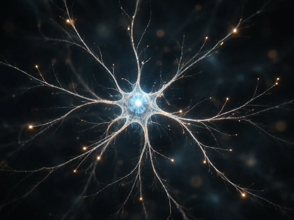
Neuronal dendrites grow because of a combination of chemical signals from the surrounding environment, electrical activity driven by experience, and internal cellular machinery that physically builds out new branches. The main triggers are neurotrophic growth factors (especially BDNF and NGF), synaptic activity that releases calcium and activates gene expression, and guidance cues that tell dendrites which direction to extend and when to stop. When any of those signals are disrupted, dendrite growth stalls, branches retract, or the whole arbor fails to form properly.
What Causes Dendrites to Grow: Signals, Mechanisms, Limits

Marcus Whitmore
30 May 2026
What dendrites actually are (and what they're not)
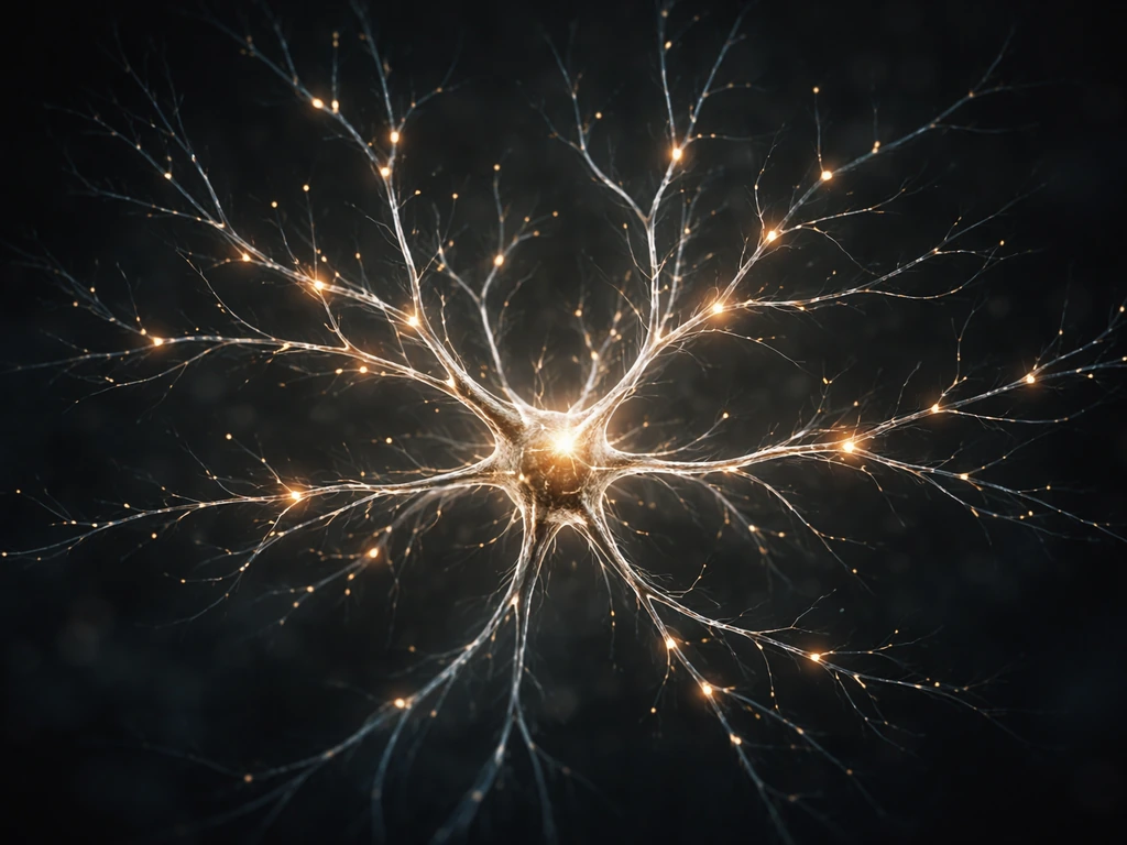
In neuroscience, dendrites are the branched, tree-like extensions of a neuron that receive incoming signals from other neurons. The word comes from the Greek for tree, which fits perfectly once you've seen a stained neuron under a microscope. Depending on the neuron type, a cell can have one dendrite or an elaborate arbor with hundreds of branches, each studded with tiny protrusions called dendritic spines. Those spines are the postsynaptic side of most excitatory (glutamatergic) synapses, and they contain dense clusters of receptors and signaling proteins called the postsynaptic density.
It's worth quickly clearing up a naming collision: in materials science, 'dendrites' also refers to branching crystal formations that grow during solidification, shaped by crystallographic anisotropy rather than anything biological. Those crystal dendrites have nothing to do with neurons. This article is entirely about the neuronal kind.
Neuron basics: structure, synapses, and why dendrite growth matters
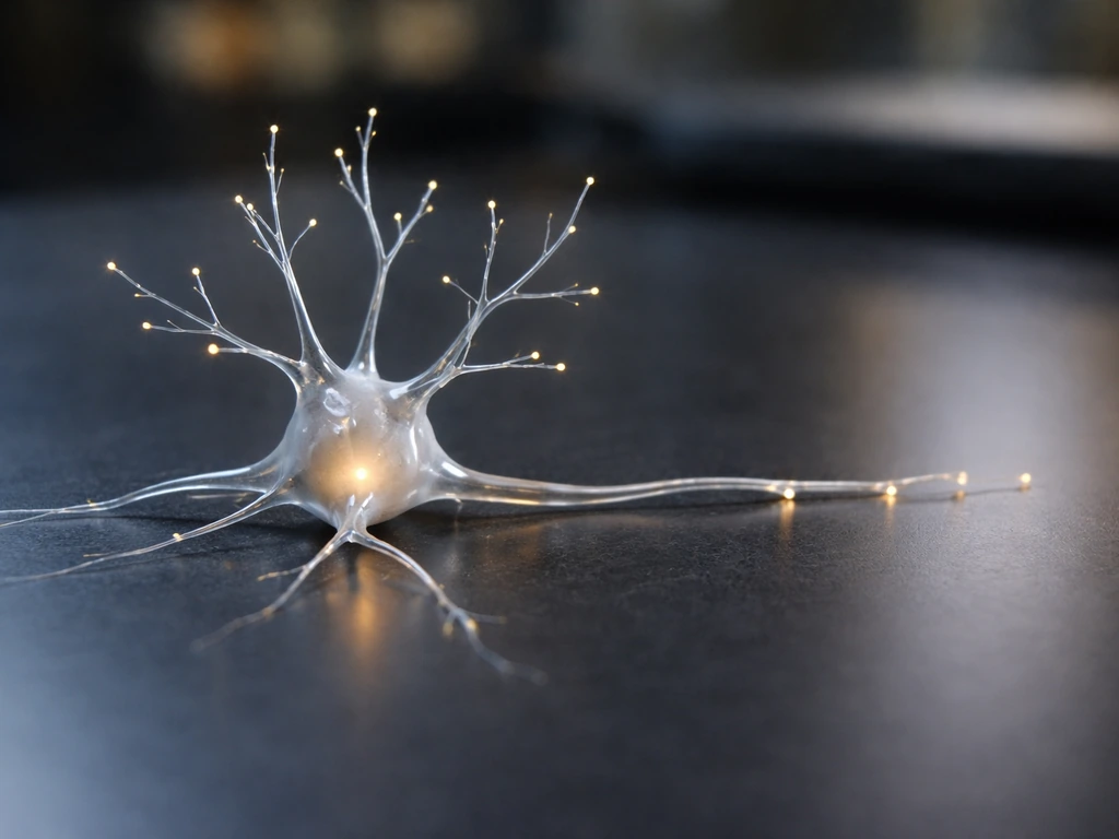
A neuron has three main compartments: the soma (cell body), the axon (which sends signals out), and the dendrites (which receive signals in). Dendrites collect synaptic inputs from potentially thousands of other neurons and integrate them before any signal propagates toward the soma and down the axon. Dendrites are branched processes associated with neurons that function to receive synaptic inputs and integrate information before signals propagate toward the soma receive signals in. So the size and complexity of a dendritic arbor directly controls how much information a neuron can take in and process.
Synapses are the connection points where one neuron's axon terminal contacts another neuron's dendrite (or spine). Excitatory synapses, which use glutamate, form asymmetric contacts with a thick postsynaptic density. Inhibitory synapses, which use GABA, form symmetric contacts and are often found directly on dendritic shafts rather than spines. The balance of excitatory and inhibitory input matters enormously for whether a dendrite grows, stabilizes, or retracts.
The cellular machinery that physically builds dendrites
Understanding what signals tell dendrites to grow is only half the story. The other half is how those signals are converted into physical branch extension. Two cellular systems do most of the heavy lifting: the cytoskeleton and membrane trafficking.
The cytoskeleton: the scaffold inside every branch
Dendrites are built on an internal scaffold made of microtubules and actin filaments. Microtubules run lengthwise through dendritic shafts and provide structural support and a track for motor proteins to haul cargo. Actin filaments dominate at the tips of growing branches and inside dendritic spines. When growth signals arrive, proteins like Rac1 and Cdc42 (small GTPases) activate actin polymerization at the branch tip, pushing the membrane outward. Microtubule stabilizing proteins like MAP2, which is actually concentrated in dendrites rather than axons, help lock in new branch length once extension occurs. Disrupting either the actin or microtubule network stops dendrite growth almost immediately.
Membrane trafficking: building material delivery
A growing dendrite needs new membrane and new proteins delivered continuously to the tip. The Golgi apparatus and endosomal recycling compartments act like a supply chain, packaging membrane vesicles and shipping them outward along microtubule tracks via motor proteins (kinesins). Local translation also plays a role: some dendritic mRNAs are transported to branch tips and translated on-site in response to synaptic activity, which allows rapid, localized addition of structural and receptor proteins without waiting for the soma to ship finished product. Disrupt vesicle trafficking or local translation and dendrite growth slows or stalls.
How neural activity and experience shape the arbor
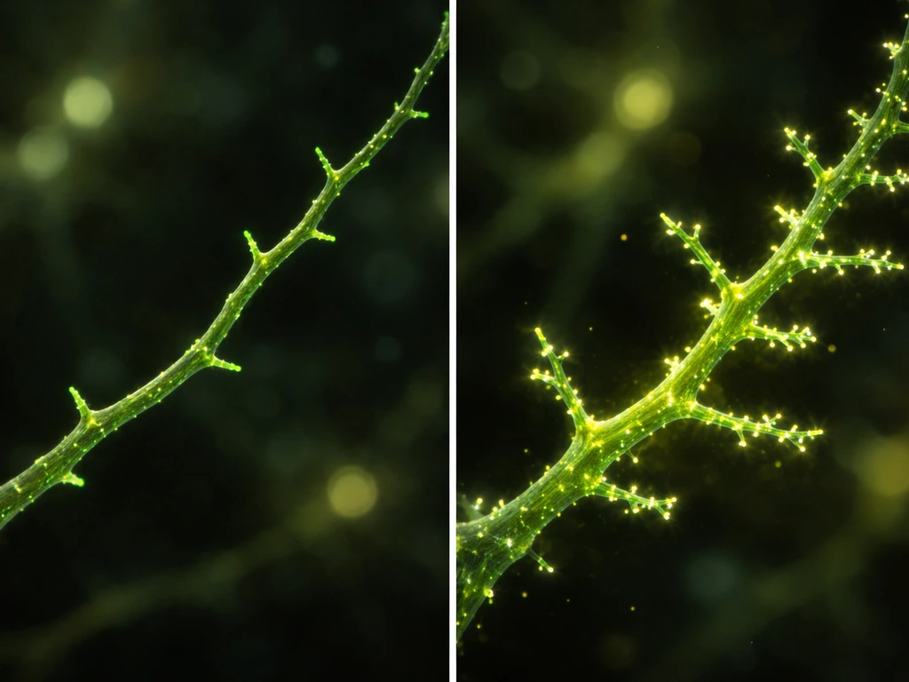
Dendrites are not static structures. They respond continuously to what the neuron experiences, which is why learning and sensory exposure physically remodel the dendritic arbor. This is the cellular basis of synaptic plasticity.
When a synapse is activated repeatedly, calcium floods into the dendritic spine through NMDA receptors and voltage-gated calcium channels. Calcium is a master second messenger: it activates kinases like CaMKII, which phosphorylates proteins that control actin dynamics, and it triggers transcription factors like CREB to switch on genes for structural growth, including genes for BDNF production. The result is spine enlargement, new branch formation, and stabilization of existing branches at active synaptic sites. Dendrites that receive no or little activity tend to retract. This 'use it or lose it' dynamic is a core feature of how experience sculpts brain connectivity, and it directly explains why enriched environments promote dendritic complexity while sensory deprivation reduces it.
This activity-dependent process is also why dendrite growth and synapse formation are tightly coupled. As dendrites grow and new synapses are formed, activity at those new synapses feeds back to stabilize the branches that host them. It's a self-reinforcing loop: activity drives growth, growth creates more synaptic surface, and new synaptic activity further stabilizes the expanded arbor.
The molecular signals that say 'grow here, stop there'
Several families of molecules act as the on/off switches and directional guides for dendrite growth.
| Signal type | Key examples | What they do |
|---|---|---|
| Neurotrophins | BDNF, NGF, NT-3, NT-4 | Bind Trk receptors; promote branching, elongation, and spine maturation |
| Attractive guidance cues | Netrin-1, Semaphorin 3A (in some contexts) | Draw dendrites toward specific target territories |
| Repulsive guidance cues | Semaphorin 3A (in other contexts), Slit, Ephrin | Prevent overlap, enforce territory boundaries, trigger retraction |
| Wnt signaling | Wnt-7a, Wnt-5a | Promotes dendrite arborization via Frizzled receptors and downstream cytoskeletal remodeling |
| Contact-dependent cues | Delta-Notch, cadherins | Stabilize specific synaptic contacts; regulate competitive branch stabilization |
| Activity-driven factors | BDNF (released by active synapses), Arc/Arg3.1 | Couple neural firing to structural growth and receptor trafficking |
BDNF deserves special attention because it appears in almost every major phase of dendrite development. During early development, BDNF released by target neurons acts as a long-range attractant. At mature synapses, locally released BDNF acts as a short-range signal that strengthens individual connections and stabilizes spines. Blocking BDNF signaling reliably reduces dendritic complexity in virtually every brain region studied.
Repulsive cues are equally important. Without molecules like Semaphorins and Slits setting boundaries, dendrites from different neurons would grow into each other's territories, creating wiring chaos. The balance between attractive and repulsive signals is what gives each neuron a characteristic, non-overlapping dendritic field.
Why dendrites don't just keep growing forever
Dendrite growth is genuinely self-limiting, and understanding the brakes is just as useful as understanding the accelerators. A few mechanisms cap expansion.
- Self-avoidance: dendrites from the same neuron express matching cell-surface proteins (like DSCAM in flies, or related molecules in mammals) that trigger repulsion when two sibling branches touch, preventing the arbor from folding back on itself.
- Competitive synapse formation: dendritic branches compete for a limited supply of presynaptic partners. A branch that fails to form stable synapses loses the activity-driven stabilization signals it needs and gets retracted.
- Resource limits: the soma can only synthesize and ship so many proteins and membrane components. As the arbor grows, metabolic demand rises; eventually resource constraints slow and then cap further expansion.
- Inhibitory feedback: as more excitatory synapses form on a growing arbor, inhibitory interneurons are recruited to balance activity. Increased inhibitory tone can suppress the calcium transients that drive further growth.
- Pruning programs: during development, genetically programmed branch pruning removes early exuberant growth to refine mature connectivity. Pruning involves localized cytoskeletal breakdown and can be triggered by signals like TGF-beta and by reduced synaptic activity.
The net result is that each neuron type reaches a characteristic mature arbor size that reflects a balance between growth signals, activity-dependent stabilization, competitive pruning, and resource availability. It's similar in principle to how a garden plant reaches a maximum canopy size when root water supply can no longer keep pace with new leaf growth.
When dendrites aren't growing: what to look for
If you're approaching this from a research, clinical, or educational angle where dendrites are failing to grow normally, the causes almost always fall into one of these categories. Checking each in order is a practical way to narrow down the problem.
- Growth factor signaling deficits: Is BDNF or another neurotrophin missing, reduced, or being blocked? Low BDNF is implicated in depression, schizophrenia, and neurodegenerative conditions. In animal models and cell culture, BDNF withdrawal reliably causes dendritic retraction within hours to days.
- Insufficient synaptic activity: Dendrites need stimulation to grow and maintain complexity. Silencing synaptic activity pharmacologically or through sensory deprivation causes arbor shrinkage. If neurons are chronically underactive (due to injury cutting off inputs, for example), expect reduced dendritic complexity.
- Excitatory/inhibitory imbalance: Too much inhibition can suppress the calcium signals that drive growth. Too much excitation causes excitotoxicity, which destroys dendrites. Both extremes are damaging. Many developmental disorders (autism spectrum conditions, fragile X syndrome) involve E/I imbalance that disrupts normal dendrite development.
- Cytoskeletal or trafficking defects: Mutations in genes for MAP2, actin regulators (like LIMK1 in Williams syndrome), or motor proteins disrupt the physical build-out of branches. These are often identifiable by abnormally short, stubby, or sparse dendrites.
- Disease and injury states: Traumatic brain injury causes rapid dendritic retraction due to calcium overload and cytoskeletal collapse. Ischemia (oxygen deprivation) destroys dendritic spines within minutes. Chronic stress reduces dendritic complexity in the prefrontal cortex and hippocampus through glucocorticoid-mediated suppression of BDNF. In Alzheimer's disease, amyloid oligomers impair synaptic function and drive spine loss before neurons die.
- Developmental timing issues: Dendrites in many brain regions follow tightly timed growth windows. If a critical signal (like a neurotrophin surge or a wave of spontaneous activity) is missed during the right developmental window, the arbor may not recover its normal complexity even if the signal is restored later.
For researchers culturing neurons, the practical checklist looks like this: confirm BDNF is present in the medium at adequate concentration (typically 25-50 ng/mL for cortical cultures), verify neurons are generating spontaneous activity (check with live calcium imaging), ensure the E/I balance isn't being crushed by excessive inhibition from immature astrocytes or contaminating inhibitory neurons, and rule out cytoskeletal damage from osmotic stress, temperature fluctuations, or phototoxicity during imaging. Those same principles can also guide practical work on how to grow neurons in a lab For researchers culturing neurons.
Putting it all together
Dendrite growth is a layered process. At the top level, neurotrophins and guidance cues tell a neuron where and how much to grow. At the middle level, synaptic activity and calcium signaling translate experience into structural change. At the bottom level, actin polymerization, microtubule stabilization, and vesicle trafficking do the actual physical work of extending branches and building spines. Each level constrains the others, which is why disrupting any one of them can stall the whole process.
Understanding how dendrites grow is inseparable from understanding how brain cells grow more broadly, and how new synaptic connections form when learning occurs. To understand how do dendrites grow in practice, it's helpful to follow how activity turns into those new synaptic connections over time. Those interconnected topics help complete the picture if you want to go deeper into any one mechanism.
FAQ
Is dendrite growth driven by one signal, or do multiple signals have to occur together?
Dendrites typically need more than one input signal at the same time. Growth factors (like BDNF/NGF), synaptic activity that elevates calcium, and permissive guidance cues must align. If one pathway is missing, the others can at best produce weak remodeling, and the branch program often fails to transition from “sprouting” to “stabilizing.”
If calcium promotes dendrite growth, why doesn’t increasing stimulation always increase dendrites?
A common lab and study mistake is treating “more calcium” as equal to “more growth.” Calcium can promote growth through kinases and gene expression, but the pattern matters (bursting versus sustained levels) and local calcium handling can differ between spines and shafts. Excessive or abnormal activity can trigger destabilization pathways, so growth outcomes depend on stimulus timing and balance.
How does the excitatory versus inhibitory balance change whether dendrites grow, stabilize, or retract?
Yes, excitatory and inhibitory inputs act like brakes and steering at the same time. Even if excitatory drive is present, too much inhibition (or too little excitatory tone) can reduce spine activation frequency, leading to retraction and fewer stabilized branches. Conversely, overly strong excitation can also harm stability by pushing plasticity programs past a beneficial range.
Are the mechanisms that extend dendrites the same as those that grow axons?
Dendrites and axons respond differently to the same internal machinery. For example, microtubule organizing and stabilizing proteins tend to be enriched in dendrites (not axons), and tip actin dynamics are especially critical for branch extension. That compartment specificity means disrupting “growth” proteins can have very different effects depending on where you target them.
How quickly do the signals that cause dendrite growth take effect, from minutes to days?
Dendrites can change rapidly at spines through synaptic activity, but long-term arbor expansion also depends on transcriptional programs and sustained availability of growth cues. That is why blocking transcription or neurotrophin signaling often reduces complexity over days, even if short-term calcium responses still occur.
What happens if local translation at dendritic tips is disrupted?
Local protein synthesis is most important when you want fast, spatially restricted remodeling at active sites. If local mRNA transport or translation is blocked, growth signaling may still be initiated, but you often get delayed or reduced spine enlargement and fewer new branches because the building blocks do not arrive efficiently at the tip.
What actually stops dendrites from growing indefinitely?
Dendrites are self-limiting because stabilization competes with removal and because resources and space become constrained as the arbor expands. In practice, competitive pruning and insufficient trophic or activity support can trim new branches before they fully mature, setting a characteristic mature complexity for each neuron type.
What if dendrites form branches but they don’t persist, what is the most likely cause?
In developmental settings, guidance cues and neurotrophins can bias initial branch placement, but activity later refines which branches persist. A useful edge case is when dendrites “appear” to grow, but they fail to stabilize because activity is missing or calcium signaling is aberrant. That can produce a network with many immature spines that later retract.
What is a practical troubleshooting path when dendrite growth stalls in culture?
For experiments aiming to promote dendrite growth in cultured neurons, it helps to monitor not just survival but spontaneous network activity and calcium dynamics, then pair that with measurements of synaptic formation and cytoskeleton integrity. If growth stalls, the next decision is usually to check whether the block is upstream (neurotrophins and cues), midstream (calcium and gene programs), or downstream (actin/microtubules and trafficking).
How do I avoid mixing up neuronal dendrite growth with “dendrites” in materials science?
One easy-to-miss issue is confusing neuronal dendrites with dendritic crystal structures in materials science. The “cause” of branching in crystals comes from physical crystallography and solidification conditions, not neurotrophins, calcium signaling, or cytoskeletal machinery, so the mechanisms are not interchangeable.
Next Article
How Do Dendrites Grow? Mechanisms and Practical Guide
How dendrites grow: outgrowth, branching, and guidance cues, plus practical ways to encourage and measure growth.