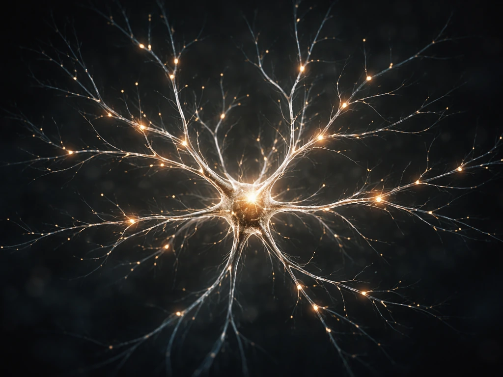
To grow neurons in a lab, you need a clean neuron source (either dissected primary tissue or stem-cell-derived precursors), a coated surface that mimics the extracellular matrix, the right media packed with survival factors, and a tightly controlled environment at 37°C with 5% CO2. Get those four things right and neurons will attach, extend neurites, and eventually wire themselves into functional networks. Skip any one of them and your cells will die within 24 hours, often without telling you why.
How to Grow Neurons in a Lab: Step-by-Step Guide

Marcus Whitmore
2 Jun 2026
Choosing your neuron source
Before you touch a pipette, decide where your neurons are coming from. That single decision shapes everything downstream: your timeline, your ethical obligations, your cost, and how closely your results mirror what happens in a living brain.
Primary neurons: fast but finite
Primary neurons are taken directly from animal embryos or early postnatal pups, most commonly rat or mouse embryonic cortex (E17-18 for cortical) or hippocampus (E18-P1 for hippocampal). They are already committed neurons, so you skip differentiation entirely. The tradeoff is that primary neurons are post-mitotic: they will not divide in your dish, they come in limited numbers per animal, and you have a narrow collection window. You also need animal use approval before you start. That said, primary cultures are the gold standard for studying basic neuronal biology because the cells behave like neurons from day one.
Stem-cell-derived neurons: scalable but slower
Induced pluripotent stem cells (iPSCs) or embryonic stem cells can be differentiated into neurons using defined chemical protocols. The appeal is scalability and, in the case of human iPSCs, direct relevance to human disease. The downside is time: full differentiation and maturation of human iPSC-derived neurons can take 4 to 12 weeks depending on the subtype, and batch variability is a real headache. If you are just starting out and your goal is to see neurite outgrowth and basic network activity, primary rodent cultures will get you there faster. If you need human neurons or genetic disease models, iPSC-derived is worth the extra work.
| Feature | Primary neurons | iPSC-derived neurons |
|---|---|---|
| Species | Rodent (mainly rat/mouse) | Human or rodent |
| Time to mature culture | 7-14 days in vitro (DIV) | 4-12 weeks |
| Proliferative? | No (post-mitotic) | Precursors divide; post-mitotic after differentiation |
| Ethical requirements | Animal use protocol (IACUC) | IRB/stem cell oversight committee |
| Batch consistency | High within a dissection | Variable; requires quality control |
| Best for beginners? | Yes | Intermediate to advanced |
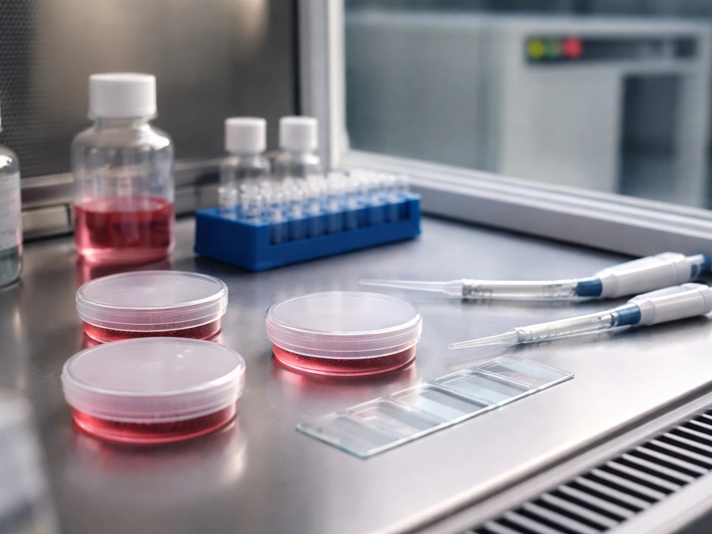
Setting up a sterile lab environment
Neurons are among the most contamination-sensitive cells you will ever culture. A yeast spore, a fingerprint, or a draft from a poorly sealed incubator door can wipe out a week's work. Everything touching your cells must be sterile: pipettes, media, tubes, and your hands (gloves, always). All cell work happens inside a biosafety cabinet (BSC) with the sash at the correct height and the cabinet running for at least 10 minutes before you open a flask.
- Wipe the BSC surface with 70% ethanol before and after each session
- Use sterile-filtered (0.22 µm) media and solutions, not autoclaved ones, for anything touching cells
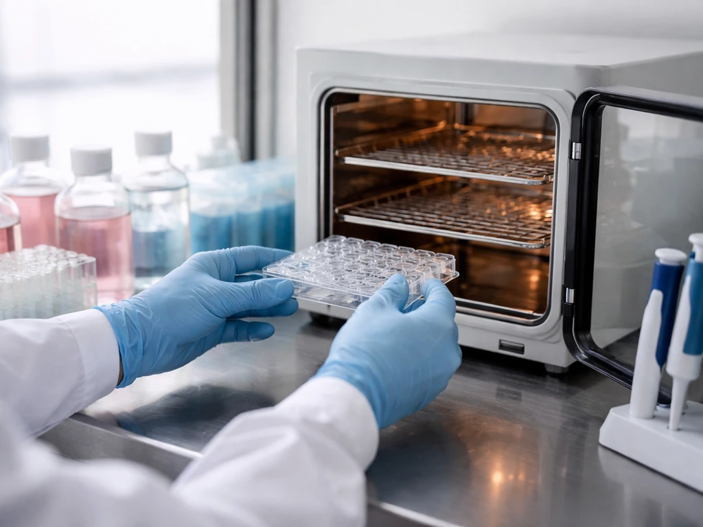
- Keep your incubator at 37°C, 5% CO2, and 95% relative humidity; check water pan levels weekly
- Work quickly but not rushed: neurons begin dying the moment they leave their normal environment
- Dedicate separate pipettes and reagents to neurons; do not share with dividing cell lines
- Add antibiotics (typically penicillin/streptomycin) to media as a contamination buffer, but treat it as a backup, not a substitute for sterile technique
One thing beginners overlook is CO2 equilibration. If you open your incubator repeatedly throughout the day, pH shifts in bicarbonate-buffered media can stress cells. Batch your media changes so you open the incubator as few times as possible.
Media, supplements, and growth factors neurons actually need
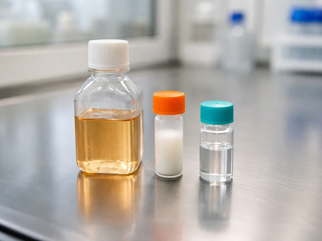
Neurons are metabolically demanding and cannot survive in generic DMEM the way a HeLa cell can. The standard backbone for primary rodent neurons is Neurobasal medium supplemented with B27 (a defined antioxidant and lipid supplement) and GlutaMAX (a stable glutamine substitute). That combination, originally optimized by Brewer and colleagues in the early 1990s, dramatically reduced glial overgrowth and extended neuronal survival compared to older serum-based recipes.
- Neurobasal + B27 (1x) + GlutaMAX (1 mM): the core recipe for most primary cortical/hippocampal cultures
- BDNF (brain-derived neurotrophic factor, 10-50 ng/mL): supports survival and promotes dendritic branching
- NGF (nerve growth factor): particularly important for peripheral and sympathetic neuron cultures
- NT-3 and NT-4 (neurotrophin-3 and -4): useful for enhancing axonal and dendritic growth in specific populations
- Cytosine arabinoside (AraC, 1-5 µM) added around DIV3-5: suppresses glial cell proliferation in primary cultures without killing post-mitotic neurons
- For iPSC-derived neurons: culture-specific factors like BDNF, GDNF, ascorbic acid, and cAMP are commonly included in maturation media depending on the subtype
Think of B27 the way you would think of soil amendments in a garden. The base medium provides water and basic salts, but without the right micronutrients and lipids, neurons starve in slow motion. Growth factors like BDNF act more like fertilizer: they do not just keep cells alive, they actively signal the cell to grow, branch, and form connections. This connects directly to how dendrites grow and why the right trophic environment is inseparable from the physical process of neurite extension.
Coating your surface: the scaffold that controls how neurons grow
A bare glass coverslip or plastic tissue culture dish is hydrophobic and biologically inert. A neuron dropped onto it will float, fail to attach, and die. You need to pre-coat the surface with molecules that mimic the extracellular matrix neurons sit on inside the brain. This is not optional. The coating you choose also directly influences neurite outgrowth, branching geometry, and how quickly synapses form.
The PDL-laminin standard
The most widely used approach for primary cortical and hippocampal neurons is a combination of poly-D-lysine (PDL) and laminin. A described poly-D-lysine (PDL) coating approach emphasizes that PDL can be used alone or in combination with laminin to improve neuronal maturation and neurite growth in primary cortical neuron cultures [A described PDL coating approach emphasizes that PDL can be used alone or in combination with laminin](https://pmc. ncbi. nlm.
nih. gov/articles/PMC10320290/). PDL (used at around 10 µg/mL) is a positively charged polymer that electrostatically attracts the negatively charged cell membrane and promotes adhesion. Laminin (used at around 5 µg/mL) is an actual extracellular matrix protein that activates integrin receptors on the neuron, triggering the internal signaling cascade that kicks off neurite extension.
After coating overnight, aspirate the solution and wash twice with sterile double-distilled water to remove unbound material before adding cells. Leaving unbound PDL on the surface can actually be toxic.
Research comparing surface coatings makes the difference concrete: laminin and Matrigel (a basement membrane extract) facilitate neuritogenesis significantly more than poly-lysine alone. If your experiment depends on measuring neurite length or branching complexity, the coating you choose will change your results, not just your survival rates.
Other coating options
- Poly-L-lysine (PLL): cheaper than PDL, widely used, but PDL is less susceptible to bacterial degradation
- Matrigel: a complex matrix extract that includes laminin, collagen IV, and entactin; excellent for neuritogenesis but batch-variable and derived from tumor tissue
- Fibronectin: good for peripheral neurons; less common for CNS cultures
- 3D hydrogel scaffolds (collagen, fibrin, or synthetic PEG-based): relevant for organoid-style cultures and studies of how physical stiffness constrains neurite growth
The physical stiffness of the substrate matters too. Neurons in the brain live in one of the softest tissues in the body, roughly 0.1 to 1 kPa. On standard rigid plastic or glass, neurons grow but their morphology and gene expression differ from what you see in vivo. For basic survival and imaging experiments, rigid coated dishes are fine. For experiments meant to model in vivo mechanics, consider compliant hydrogel substrates.
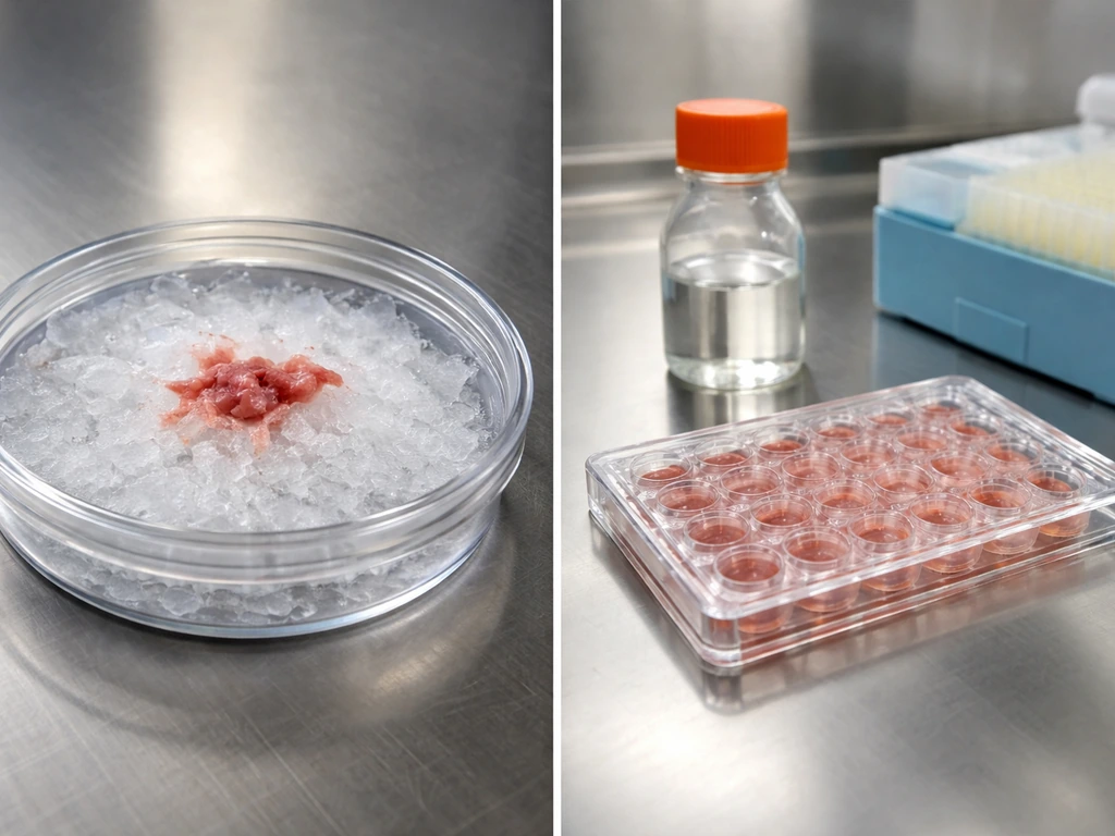
Seeding, differentiation timelines, and keeping cultures alive
Primary neuron seeding and early days
For primary dissociated cortical or hippocampal neurons, typical seeding densities range from 50,000 to 200,000 cells per cm2 depending on your application. High-density cultures (100,000-200,000/cm2) form robust networks with spontaneous activity and are good for electrophysiology. Low-density cultures (25,000-50,000/cm2) let you image individual neurons and trace complete morphology but require more careful maintenance. Seeding too densely causes nutrient depletion and waste accumulation faster than you can exchange media, which mirrors a broader principle: growth in any system is ultimately constrained by resource diffusion and waste removal.
After plating, leave cells undisturbed for at least 4 hours, ideally overnight, before any media change. They need time to settle and attach. Your first half-media change at DIV1 removes dead cells and debris without disturbing the ones that have just anchored. From there, change half the media every 2-3 days. Never do a full media change: the conditioned media contains secreted factors (including neurotrophins and glutamate receptor modulators) that the neurons themselves produce, and wiping them out sets the culture back.
iPSC differentiation and maturation timeline
For iPSC-derived neurons, the workflow has more stages. A typical cortical differentiation protocol proceeds through: (1) neural induction using dual SMAD inhibition (days 0-10) to convert pluripotent cells to neural progenitor cells (NPCs), (2) NPC expansion and patterning (days 10-25) where growth factors specify regional identity, and (3) terminal differentiation and maturation (weeks 4-12) where progenitors exit the cell cycle and become functional neurons. Because neurons are post-mitotic once differentiated, you cannot passage them like you would a stem cell line. Plan your experiment so you plate differentiated neurons at the density you need from the start.
Media change rhythm and nutrient constraints
A key biological constraint in any culture system is the limit on nutrient diffusion. In a dish, neurons at the bottom have to extract oxygen and glucose from a thin layer of media sitting above them. As cultures mature and metabolic demand increases, waste products like lactate and glutamate accumulate. Stale media smells faintly acidic and turns yellow from pH-sensitive phenol red. At that point, neurons are already stressed. Consistent half-media changes every 2-3 days keep concentrations in a livable range, but they are a workaround for what the body solves with blood flow.
Measuring actual growth: beyond just counting live cells
A neuron that is alive is not necessarily a neuron that is growing. To know whether your culture is working, you need multiple readouts across viability, morphology, and function.
Viability
- Live/dead staining: calcein-AM (green, live) and ethidium homodimer-1 (red, dead) give a quick visual snapshot
- MTT or resazurin (alamarBlue) assays: plate-reader-based metabolic activity measurements; good for quantifying survival across conditions
- Phase contrast microscopy: healthy neurons have bright, phase-bright cell bodies with visible processes; dead neurons are shrunken, dark, or swollen
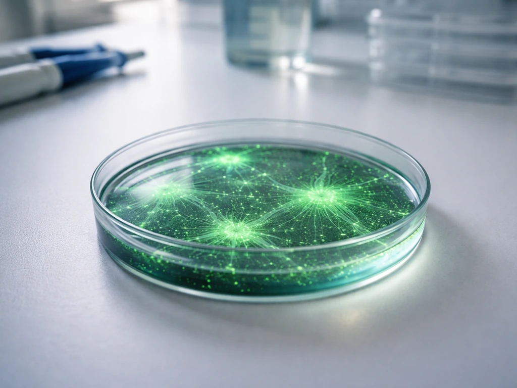
Neurite outgrowth and dendritic growth
Neurite length and branching are your primary morphological readouts for growth. Fix cells at your chosen time point with 4% paraformaldehyde, then stain with MAP2 (a dendritic marker) and Tuj1 or beta-III-tubulin (marks all neuronal processes). Imaging these under a fluorescence microscope and tracing processes with software like ImageJ/FIJI with the Simple Neurite Tracer plugin gives you quantitative data on total neurite length, number of branches, and Sholl analysis (a measure of branching complexity as a function of distance from the cell body). This is the same growth process discussed in how dendrites grow and what drives their branching patterns.
Synaptic markers
Around DIV14-21 in primary rodent cultures, synapses begin forming in earnest. Stain for pre-synaptic markers (synapsin-1, synaptophysin, vGlut1) and post-synaptic markers (PSD-95 for excitatory synapses; gephyrin for inhibitory). When you see punctate pre- and post-synaptic staining that co-localizes along dendrites, you have structural synapses. That is a big milestone.
Functional readouts
- Calcium imaging with GCaMP (genetic indicator) or Fluo-4 (chemical dye): spontaneous calcium transients indicate network activity without any hardware beyond a fluorescence microscope and camera
- Multi-electrode arrays (MEAs): culture neurons directly on electrode arrays to record extracellular spikes and bursting patterns; this is the most accessible electrophysiology for beginners
- Patch-clamp electrophysiology: the gold standard for single-cell functional assessment, but requires specialized equipment and significant training
What goes wrong and how to fix it
Most neuron culture failures trace back to a small set of root causes. Here is what to look for and where to start when things go sideways.
| Problem | Likely cause | Fix |
|---|---|---|
| Mass cell death within 24-48h | Poor dissociation, coating failure, or contaminated media | Check coating protocol; re-filter media; verify incubator CO2 and temperature |
| Cells attach but no neurites by DIV3 | Missing growth factors, wrong coating, or overly rigid substrate | Add laminin to coating; confirm BDNF is in media; check supplement lot |
| Glial overgrowth by DIV7-10 | Missing AraC treatment or too high seeding density | Add AraC (1-5 µM) at DIV3-5; reduce seeding density |
| Low plating efficiency (cells don't stick) | Coating was not done correctly or was washed off | Re-coat; ensure PDL/laminin incubation was overnight; rewash with ddH2O not PBS |
| Rapid media acidification (yellow within 1 day) | Too many cells, contamination, or excess glutamate | Reduce density; check for contamination; switch to GlutaMAX if using L-glutamine |
| Batch-to-batch variability in iPSC cultures | Inconsistent differentiation efficiency or cell passage number | Use quality-controlled NPC stocks; track passage number; verify neural identity by flow cytometry before plating |
| Contamination (cloudy media, fuzzy growth) | Non-sterile technique or reagents | Discard affected cultures; deep-clean incubator; re-examine sterile technique step by step |
One failure mode that surprises beginners is glial overgrowth. In primary cultures, you inevitably plate some glia alongside neurons. If you skip AraC or use serum-containing media, glia proliferate aggressively and physically crowd out neurons by DIV10-14. This is a density-dependent constraint: glia sense neighbor cells via contact inhibition, but in rich media they override it. AraC selectively targets dividing cells (glia) while sparing post-mitotic neurons.
Safety, ethics, and picking a real starting protocol
Biosafety and compliance
Primary rodent neuron work requires an active animal use protocol approved by your institution's IACUC (Institutional Animal Care and Use Committee) before you collect any tissue. Human iPSC-derived neurons require IRB oversight if cells are derived from human donors, plus approval from your institution's stem cell research oversight (SCRO) committee. Do not assume you can start without these approvals. The paperwork exists to protect the animals, the human donors, the researchers, and the integrity of the science.
- Work at BSL-1 or BSL-2 depending on your cell source and any viral vectors used (e.g., AAV or lentivirus for transfection moves you to BSL-2)
- Dispose of all biological waste in autoclave bags or approved biohazard containers; never pour cell-containing media down a regular drain
- Formaldehyde and paraformaldehyde (used for fixation) are hazardous; use in a fume hood and dispose of as chemical waste
- Document every culture: date, cell source, passage or dissection number, coating lots, and media lots; troubleshooting is nearly impossible without records
Your practical starting point today
If you are setting up for the first time, start with rat embryonic cortical neurons (E17-18). They are robust, well-characterized, and there are dozens of published protocols to compare against. The PDL-laminin coating with Neurobasal/B27/GlutaMAX media is the most replicated baseline in the field. Use it exactly as written in at least one published protocol before you start optimizing. Changing multiple variables at once is the fastest way to not know why something failed.
For iPSC beginners, commercial differentiation kits from suppliers like Thermo Fisher (Gibco PSC Neural Induction Medium) or StemCell Technologies give you a reproducible starting point with technical support. They are more expensive than making your own media from scratch, but the reduction in variability is worth it while you are learning the system.
Once your baseline culture is working and you can reliably see neurite outgrowth at DIV7 and synaptic puncta at DIV14-21, you will have built the foundation to explore more complex questions: how specific genes regulate how brain cells grow, what causes dendrites to branch or retract, or how network-level activity emerges from individual synaptic connections. If you are wondering how brain cells grow, focus on the signals that drive neurite extension, branching, and maturation over time. That is where the real biology starts.
FAQ
How can I tell early on whether my neurons are attaching versus just surviving?
Attachment should show up as neurons settling and extending processes within the first 4 to 24 hours, not just remaining viable. A quick check is to image at the same field and exposure across days 0 and 1, then compare neurite emergence and cell flattening on the coated surface. If cells look rounded and do not extend processes by around DIV1 to DIV2, it often indicates a coating or plating issue rather than a media problem.
Do I need to use conditioned media from an existing culture, or can I rely only on my standard supplements?
For primary cultures, half-media changes are designed to preserve neuron-secreted factors already in your dish, so you typically do not need external conditioned media. The exception is when you are adapting a new batch of cells to a new subtype or coating, where a short acclimation with a small amount of conditioned media can help. If you do this, keep the percentage low and consistent so you can still interpret experimental effects.
Why do neurons sometimes die after a media change, even if I changed at the right days and frequency?
Most media-change deaths come from mechanical disturbance, temperature/pH shock, or leaving cells out of the incubator too long. Use pre-warmed media, work efficiently, and avoid directly blasting neurons during aspiration. If you must troubleshoot, standardize timing and handling length, and ensure your pipette tip is positioned near the edge of the well rather than over the cell layer.
Can I grow neurons in serum instead of the defined Neurobasal/B27/GlutaMAX approach?
You can, but serum increases glial proliferation and can change neuronal maturation and baseline excitability, which makes both morphology and network readouts harder to interpret. If you use serum-containing media, you generally need additional strategies to control glia and you should not compare directly to results obtained with fully defined media recipes.
What’s the difference between neurite outgrowth problems caused by density versus coating?
Low density often yields sparse networks with limited neurite intersections, while coating failures usually produce poor attachment, drifting cells, and minimal neurite initiation across most cells. A practical decision rule is to compare multiple densities on the same coated batch. If attachment is poor at all densities, it points to coating chemistry, coating concentration, or wash/removal steps.
How do I handle CO2 and pH if my incubator is shared or I frequently check cultures throughout the day?
Minimize door openings as described, but also reduce the time culture plates spend outside the incubator. Use a consistent workflow order across dishes, and batch all imaging and media prep. If you suspect pH swings, monitor with your media color change trends across the same incubation schedule, then standardize handling time before changing experimental variables.
Is it better to do full media changes at some point, for example when waste builds up?
For most neuron cultures, full replacements increase disturbance and remove protective soluble factors. Instead of switching to full changes, extend or tighten the half-media schedule based on culture health. If phenol-red color shifts rapidly or cells show stress signs, that’s a cue to adjust timing of half changes, not to wipe out all conditioned components at once.
How do I choose seeding density when my endpoint is network activity versus single-cell morphology?
Higher density (roughly 100,000 to 200,000 cells per cm2) tends to improve spontaneous activity and synaptic connectivity, which is useful for electrophysiology and calcium imaging. Lower density (about 25,000 to 50,000 cells per cm2) usually helps you trace individual morphologies and quantify neurite complexity per neuron. If you are unsure, run a small density pilot on the same day to confirm which range gives you reliable branching metrics and detectable activity.
What should I do if I see glial overgrowth despite using a defined media recipe?
First, verify whether you included a glia-limiting step such as AraC when your protocol calls for it, and confirm it was applied at the correct timing window. Next, check plating density and whether you are using serum or adding supplements that may favor glial division. Finally, evaluate your starting preparation, since an overly glia-rich dissociation or insufficient separation can overwhelm even good downstream conditions.
How can I avoid batch-to-batch differences when differentiating iPSC-derived neurons?
Batch variability usually comes from starting cell quality, differentiation stage timing, and how strictly you follow the induction and patterning windows. Use consistent handling and passage history for the pluripotent cells, record day-by-day markers or checkpoints, and plate differentiated neurons at a fixed density from the same differentiation batch. If your endpoint is functional, consider running more than one differentiation batch before committing to a key experiment.
Are neuron cultures compatible with high-throughput imaging setups, and what usually breaks first?
They can be compatible, but the most common failure is increased handling time and temperature exposure during plate swaps and autofocus cycles. Standardize plate preparation so cultures spend minimal time outside the incubator, and confirm that your coating and well type are consistent across plates. Also, track whether your imaging volume and light exposure affect cell health over multiple time points.
What’s a practical way to decide when to fix and stain for neurite and synapse assays?
Use time windows rather than a single day. For neurites, confirm that outgrowth is present and increasing around DIV7, then pick a consistent endpoint for quantification (for example, when branching is clearly established). For synapses, schedule structural staining around DIV14 to DIV21 in primary cultures and ensure you are imaging comparable regions and using the same thresholding approach so puncta counts are comparable across wells.
Next Article
What Causes Dendrites to Grow: Signals, Mechanisms, Limits
Learn what causes dendrites to grow: signals, cytoskeleton and transport, neural activity, and limits on branching.