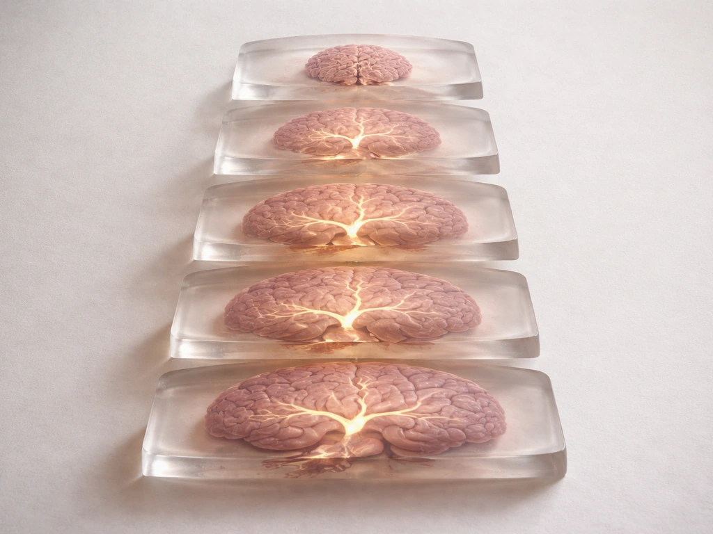
When dendrites grow and new synapses are formed, your brain is physically rewiring itself. A dendrite sprouts a tiny protrusion called a spine, that spine makes contact with a neighboring axon, and within roughly 45 minutes the two sides start recruiting the molecular machinery that turns a casual touch into a real, working synapse. This isn't metaphor, it's measurable structural change that happens during learning, enriched experience, and even in old age under the right conditions.
Dendrites Grow and New Synapses Are Formed: Mechanisms

Marcus Whitmore
7 May 2026
What dendrite growth and synapse formation actually mean
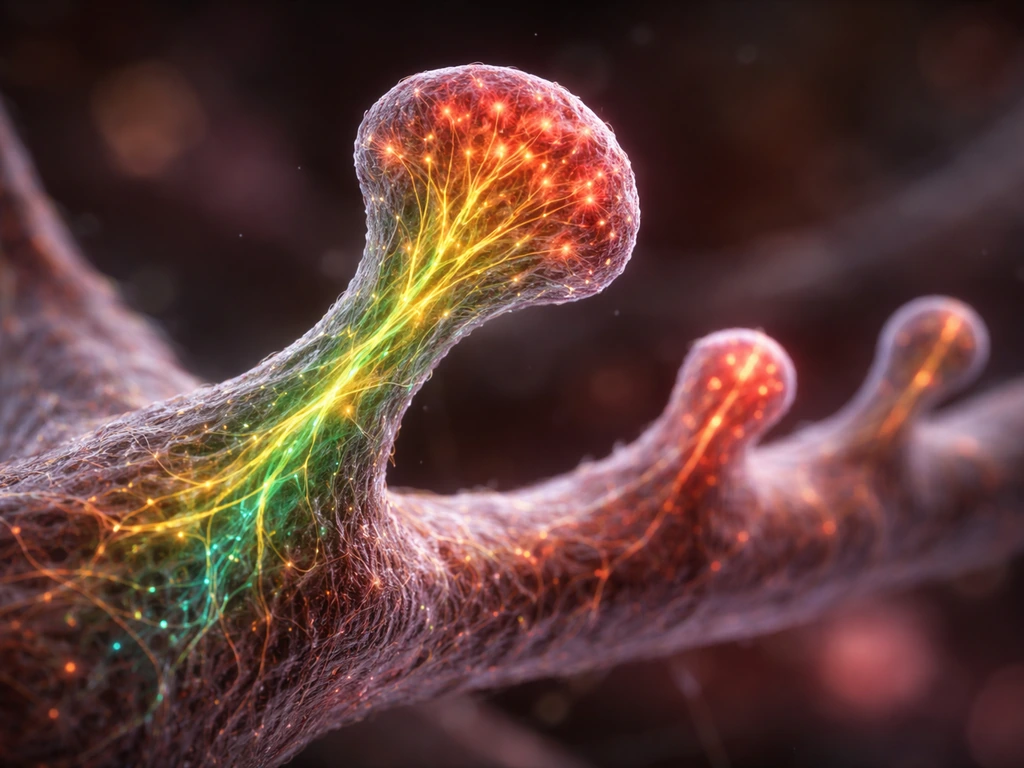
Dendrites are the branching input arms of a neuron, think of them like the antennae on a satellite dish, tuned to catch signals from other cells. When we say dendrites 'grow,' we usually mean one of a few distinct things: the overall arbor (the tree-like branching pattern) gets more complex, the total length of branches increases, or small protrusions called dendritic spines increase in number or size. These are not the same event, and they don't always happen together.
A synapse is the functional connection point between two neurons, one presynaptic (the signal sender) and one postsynaptic (the receiver). Synapse formation, or synaptogenesis, is the process by which a new junction is built, complete with a presynaptic active zone full of vesicles on one side and a postsynaptic density (PSD) packed with receptors on the other. You can have a dendrite that grows without forming synapses, and you can have synaptic strengthening without obvious structural growth. Most of the exciting biology happens when both occur together.
The cellular machinery driving dendritic remodeling
The skeleton inside a dendritic spine is made almost entirely of actin filaments, and the whole game of spine growth and shrinkage is really a game of actin dynamics. When a spine enlarges, actin polymerizes outward. When it retracts, actin disassembles. The key molecular players controlling this are the Rho-family GTPases, a group of molecular switches including Rac1, Cdc42, and RhoA. Blocking Rac or Cdc42 reduces dendrite branching in developing neurons, while activating them promotes spine growth. In a stimulated spine, Cdc42 activity stays tightly local while activated Rac1 and RhoA spread about 5 micrometers out into the dendritic shaft, this compartmentalization is how the cell targets remodeling to specific sites without accidentally reshaping everything nearby.
Rac1 and Cdc42 drive spine growth by activating the WAVE regulatory complex and N-WASP, which in turn activate the Arp2/3 complex to build branched actin networks. Rac1 activation also connects to NMDA receptor signaling through a WAVE complex component called Cyfip1, which is a direct mechanistic link between synaptic electrical activity and physical spine shape change. PAK kinases (including PAK3) sit further downstream and translate Cdc42/Rac signals into rapid cytoskeletal reorganization. If you want to understand dendritic spine biology at a mechanistic level, the Rho GTPase-actin-Arp2/3 axis is the core circuit worth knowing.
Scaffold proteins also shape the dendrite from the inside out. Homer1b/c, for example, forms a mesh-like matrix that anchors Shank proteins and actively promotes spine growth and morphology. The activity-inducible form Homer1a works differently, it competes with the long forms and can disrupt or reshape the scaffold, which is part of how acute activity reconfigures synaptic structure on a fast timescale.
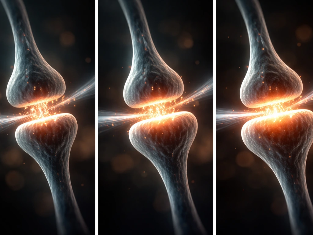
How synapses start: formation, recruitment, and stabilization
The assembly of a new synapse follows a surprisingly orderly sequence. In cultured hippocampal neurons, the presynaptic side gets its act together first: active zone proteins like Bassoon cluster at a new bouton, and only about 45 minutes later does the postsynaptic density protein PSD-95 (and glutamate receptors) appear on the dendritic side. This temporal order matters because it tells you the presynaptic terminal is largely self-organizing and 'invites' the postsynaptic response rather than both sides building simultaneously.
The molecular handshake across the synapse is managed by adhesion molecules, most famously the neurexin-neuroligin pair. Neurexins sit on the presynaptic membrane; neuroligins sit on the postsynaptic side. They bind each other across the synaptic cleft and trigger differentiation on both sides. Purified neuroligin alone is enough to cluster neurexin and induce presynaptic differentiation in culture experiments. Neuroligin family members (NLGN1, NLGN2, NLGN3) can drive both excitatory and inhibitory synapse formation depending on their partners. N-cadherin adds to this by stabilizing the synapse over the long term, particularly during and after plasticity events.
Once the initial contact is made and the scaffold assembles, the synapse needs to be maintained. PSD-95 accumulates in a protracted, asynchronous way over hours to days in new spines. The synaptic tag-and-capture model proposes that local activity creates a molecular 'tag' at a synapse, and plasticity-related proteins (PRPs) synthesized in the cell body or locally in dendrites get captured selectively at tagged sites. Arc/Arg3.1 mRNA is one such locally trafficked molecule, it is synthesized near recently active synapses and plays a role in consolidating plastic changes.
Activity-dependent plasticity and learning signals
The classic principle here is Hebbian: neurons that fire together, wire together. The molecular implementation runs through NMDA receptors, which act as coincidence detectors, they open only when glutamate is present AND the postsynaptic membrane is already depolarized. When both conditions are met, calcium floods in, activating CaMKII (calcium/calmodulin-dependent protein kinase II). CaMKII then phosphorylates AMPA receptors and promotes their insertion into the synapse, which is the core mechanism of long-term potentiation (LTP), the most studied cellular correlate of learning.
Spike-timing-dependent plasticity (STDP) adds precision: if a presynaptic neuron fires just before the postsynaptic one (within a window of roughly 10–20 ms), the synapse strengthens (LTP). If the order is reversed, it weakens (LTD). A standard STDP induction protocol uses 60–100 repetitive pairings at 0.2–5 Hz, and the outcome flips depending on that millisecond timing window. STDP rules also shift depending on where on the dendrite the synapse sits, distal inputs can actually show LTP with post-before-pre pairings when combined with high-frequency bursts, which makes dendritic location a real variable in plasticity outcomes.
The brain also runs a parallel homeostatic system. If a neuron is chronically silenced (e.g., by the sodium channel blocker tetrodotoxin for 3–4 days), it multiplicatively scales up the amplitude of all its miniature excitatory postsynaptic currents (mEPSCs), a process called synaptic scaling. This is the brain's way of keeping its overall excitability tuned. Importantly, homeostatic scaling also changes dendritic spine volumes and shifts the threshold for subsequent Hebbian plasticity, so the two systems are not independent.
At the gene expression level, activity triggers immediate-early genes like Arc/Arg3.1, regulated through CREB, MEF2, SRF, and ERK signaling pathways, as well as Egr transcription factors (Egr1 and Egr3) that directly bind the Arc gene promoter. Arc protein is then transported to recently activated synapses, linking nuclear transcription to local synaptic remodeling in a spatially precise way.
Development vs adulthood: what changes across the lifespan
During early brain development, synaptogenesis runs at a massive scale, the human cortex overproduces synapses in the first years of life and then prunes them back. Microglia are central to this pruning process, engulfing synaptic elements using complement cascade proteins (C1q and C3) as 'eat me' signals. Mice lacking C3 or its receptor CR3 fail to properly refine their retinogeniculate connections, showing large, persistent defects in circuit wiring. The microglial receptor CX3CR1 is also implicated in spine elimination during these critical periods. This means the developing brain is simultaneously making and removing synapses, with the balance shaped by sensory experience.
In the adult brain, the rules shift but the game continues. Adult hippocampal CA1 spines change on the timescale of days during learning and environmental enrichment, as shown by in vivo longitudinal imaging. In adult primates, a month in a complex environment increased dendritic tree length and complexity, spine density, and synaptic protein levels in the hippocampus and prefrontal cortex. In rats, environmental enrichment raised spinophilin mRNA (a spine marker) for up to four weeks. Even in the oldest-old rats, enriched housing increased dentate granule cell dendritic tree complexity by 61% (total segment number) and 116% (length), with spine density rising 88%. The adult brain is not structurally static, it is just slower and more selective than the developing brain. Roughly 40% of spines in sensory and motor cortex turn over every five days under normal conditions.
Adult hippocampal neurogenesis adds another layer: new neurons born in the dentate gyrus actually go through synaptogenesis themselves, integrating into existing circuits with synaptic ultrastructure typical of mature granule cells. This process is sensitive to enrichment, exercise, and stress, making it one of the more tractable targets for understanding adult synaptogenesis.
What limits dendrite growth and synapse formation

Growth doesn't happen in a vacuum, and there are real constraints worth understanding if you're trying to promote or study this process.
- Inhibitory molecules in myelin: Adult CNS myelin contains Nogo, MAG, and OMgp, which bind the Nogo-66 receptor complex (NgR with co-receptors p75/TROY and LINGO1) and suppress neurite outgrowth. These don't affect dendritic spine turnover in intact circuits the way they block axon regeneration, but they're a hard wall for long-distance growth.
- Extracellular matrix barriers: Chondroitin sulfate proteoglycans (CSPGs) in the extracellular matrix, particularly around perineuronal nets, restrict structural plasticity in adult cortex. Degrading CSPGs with chondroitinase reopens critical-period-like plasticity in animal models.
- Metabolic limits: Dendrite growth and synapse formation are energetically expensive. Local protein synthesis in dendrites requires ribosomes, mRNA trafficking, and ATP. Oxygen and glucose supply at the level of individual dendrites (supported by the surrounding vasculature) sets a real ceiling on how much remodeling can happen.
- Competition for trophic support: Neurons compete for neurotrophic factors like BDNF and NGF. BDNF-TrkB signaling promotes both dendritic growth and synapse stabilization, but the supply isn't unlimited. Spines that don't receive sufficient trophic signaling get pruned.
- Wiring rules and circuit identity: Not every axon-dendrite contact becomes a synapse. Molecular identity (matching receptor/ligand pairs like specific neurexin-neuroligin combinations) determines whether a contact matures. A neuron won't form a synapse with a mismatched partner just because they touch.
How to promote it or study it: practical next steps
Whether you're designing a learning intervention or running a neuroscience experiment, the mechanisms above translate into concrete approaches. Researchers also study how neurons are grown in a lab by manipulating conditions that affect dendrite development and synapse formation. Here's how to think about both.
For learning and cognitive enrichment
Environmental enrichment in animal studies combines novelty, physical activity, and social interaction, and each of those components contributes independently. For humans, the closest translation is combining genuine cognitive challenge (new skills, not just repetition), aerobic exercise (which robustly upregulates BDNF), and social engagement. Spaced repetition hits the STDP-relevant timing window better than massed practice. Sleep matters enormously for consolidation, as it's the window when many plasticity-related proteins are synthesized and synaptic tags are resolved. The key is that passive exposure doesn't drive structural change nearly as well as active, demanding engagement with new material.
For experimental measurement
If you're studying synaptogenesis in the lab, these are the standard readouts and what they tell you:
| Method | What it measures | Key markers / details |
|---|---|---|
| Confocal immunofluorescence | Synapse density and co-localization | PSD-95 (postsynaptic) + synaptophysin or Bassoon (presynaptic); colocalization fraction rises from ~37% at 8 DIV to ~75% at 14 DIV in cortical cultures |
| 2-photon in vivo imaging | Spine turnover and structural dynamics in live animals | Thy1-GFP or similar sparse labeling; track formation/elimination rates over days to weeks |
| Electrophysiology (whole-cell patch) | Functional synapse strength and number | mEPSC frequency (synapse number proxy) and amplitude (single-synapse strength); use TTX for homeostatic scaling paradigms |
| STDP induction protocol | Synapse-specific LTP or LTD | 60–100 pairings at 0.2–5 Hz; vary pre/post timing window (±10–50 ms) to probe directionality |
| Immediate-early gene readouts (Arc, Homer1a) | Recent activity and plasticity events | In situ hybridization or Arc-GFP reporter mice; Arc mRNA localizes to recently activated dendrites within minutes |
| Super-resolution imaging (STORM/PALM/STED) | Nanoscale organization of PSD molecules | GluA2 surface expression, PSD-95 cluster size; sensitive enough to detect homeostatic scaling of individual synaptic zones |
| Western blot / qPCR | Population-level protein and mRNA changes | Spinophilin, PSD-95, synaptophysin, Arc, BDNF; useful for enrichment studies or drug treatment timecourses |
For a clean experimental pipeline, start with immunofluorescence colocalization (PSD-95 + Bassoon or synaptophysin) to get a synapse count, add electrophysiology to confirm function, and use an IEG reporter to tag recently active synapses. Time-lapse imaging of PSD-95-GFP lets you watch new synapses assemble in real time, you can literally see PSD-95 puncta appear and grow over hours in cultured neurons.
Safety and ethical caveats
It's worth being direct here: the mechanisms described above are well-established in animal and cell culture models, but translating them into 'brain enhancement' in humans requires serious caution. Drugs that block myelin inhibitors, degrade CSPGs, or amplify NMDA receptor signaling can also lower seizure thresholds, disrupt circuit specificity, or cause excitotoxicity if dosed incorrectly. BDNF itself has poor blood-brain barrier penetration and complex dose-response relationships in vivo. Exercise, enriched environments, sleep optimization, and spaced learning remain the safest and most evidence-grounded interventions available to most people today. For researchers, all in vivo animal work requires appropriate institutional approval, and claims about 'enhancing' human cognition through pharmacological plasticity manipulation are far ahead of the current evidence base.
If you want to go deeper on the upstream biology, how dendrites physically extend their arbors in the first place, what molecular signals drive dendritic outgrowth during development, or how neurons are born and integrated in adult tissue, those mechanisms build naturally on everything covered here and are worth exploring as the next layer of this topic. how do brain cells grow how dendrites physically extend their arbors in the first place. At a mechanistic level, it's useful to compare these synaptic and remodeling signals with how do dendrites grow during development. Those upstream signals and pathways are what cause dendrites to grow during development and remodeling.
FAQ
Does dendrite growth always mean new synapses are being formed?
Not necessarily. Dendritic growth (more branching, longer branches, or more spines) can occur without forming functional synapses, and conversely synapses can strengthen while spine size and number stay relatively stable. A practical test is to pair a structural readout (for example, PSD-95 with a presynaptic marker) with a functional readout (electrophysiology or calcium imaging) rather than relying on spine counts alone.
Why might I see dendritic or bouton changes but not postsynaptic density in synaptogenesis experiments?
Timing is a real experimental variable. Presynaptic components often appear before the postsynaptic density in developing synapses, so if you only measure one timepoint you can miss either the initial contact phase or the later receptor recruitment phase. Plan a short time series around the expected window (for example, hours in culture) and interpret “no PSD-95” at early times as a possible delay rather than a failure of synaptogenesis.
Can synapses strengthen without obvious changes in spine structure?
Yes, synaptic and structural plasticity can partially decouple because different steps have different rate limits. For example, glutamate receptor insertion and phosphorylation can occur without obvious actin remodeling over the same measurement window, while spine enlargement depends strongly on actin dynamics and local signaling. To avoid false conclusions, use parallel readouts: receptor function or phosphorylation plus spine morphology and actin-based markers.
How does synaptic scaling affect experiments looking for LTP or STDP?
Homeostatic scaling can shift where a neuron “starts” for future Hebbian plasticity by changing excitability and synaptic thresholds. So an intervention that changes network activity may alter LTP or LTD rules indirectly even if the Hebbian machinery (NMDA, CaMKII, AMPA insertion) is intact. If you are testing STDP or LTP, verify baseline activity levels and miniature event properties before and after your manipulation.
Why doesn’t my STDP experiment match the expected LTP-for-pre-before-post rule?
STDP outcomes depend on both timing and protocol details. In particular, the induction window and the firing pattern (including burst structure) can flip the sign of plasticity for certain dendritic locations. If your results disagree with a “standard” STDP rule, check whether your neurons show realistic burst timing and whether the recorded synapses are proximal versus distal.
Does the location of a synapse on the dendrite change whether it strengthens or weakens?
Spine localization within the dendritic tree changes effective signaling. Distal synapses experience different calcium dynamics, attenuation, and local compartmentalization, so plasticity can differ even with the same presynaptic and postsynaptic spike timing. When possible, restrict analysis to a defined dendritic distance range, or include dendritic geometry as a factor in interpretation.
If neuroligin drives synapse formation, can it predict whether the synapse becomes excitatory or inhibitory?
Yes, neuroligin family members can bias whether the connection matures into excitatory or inhibitory forms, but the final outcome also depends on the cellular context and the matching presynaptic partners. In practice, if you induce or perturb adhesion molecules, you should confirm synapse type with functional assays (for example, excitatory versus inhibitory receptor currents) rather than assuming adhesion specificity alone determines excitatory versus inhibitory identity.
Why do functional changes sometimes appear before PSD-95 puncta become obvious?
Actin remodeling often limits how quickly spine morphology changes, while PSD accumulation and receptor trafficking can follow later, asynchronously. A common mistake is to treat the time course of PSD-95 appearance as identical to the time course of functional strengthening. Use a schedule that includes early molecular events (for example, receptor or signaling readouts) plus later structural/PSD readouts.
What is the most common reason enrichment does not produce dendrite or spine changes?
Environmental enrichment works by multiple partially independent components, and “newness” matters. Passive exposure or repetition without challenge often produces weaker structural effects because it does not robustly engage the coincidence detectors and activity-dependent transcriptional programs needed for lasting remodeling. For translation or intervention design, build in cognitive novelty, not just more practice time.
Why is it risky to try to “boost synapses” by amplifying NMDA signaling broadly?
Not in a simple linear way. Enhancing global NMDA signaling or broadly suppressing inhibition can increase seizure risk or excitotoxicity, even if plasticity signatures look more “active.” For safety and interpretability, consider dose and specificity, monitor network stability, and treat “more plasticity” as potentially maladaptive if it disrupts proper circuit tuning. This is especially important for any claim about human cognitive enhancement via pharmacology.
What are the biggest mistakes when quantifying dendritic spines and synapses in vitro?
Common lab pitfalls include counting spines without confirming they correspond to synapses, using only one marker that can be present during immature stages, and ignoring whether the synapse is pre- and postsynaptically apposed. A robust pipeline combines colocalization with a presynaptic marker and a postsynaptic scaffold (for example, Bassoon plus PSD-95), and verifies function with electrophysiology or live activity reporters.
Do the mechanisms for dendrite growth and new synapse formation work the same way in development versus adulthood?
Yes. If you are manipulating development or adult plasticity, you can change baseline scaffold composition, cytoskeletal state, and receptor trafficking capacity, which alters how the same upstream signals translate into growth and synapse maturation. A decision aid is to include an age-matched control and measure both morphology (spines, arbor metrics) and molecular state (for example, actin dynamics or PSD components) rather than assuming one mechanism scales across ages.
Next Article
How Do Brain Cells Grow From Birth to Adulthood
How brain neurons and glia grow from birth to adulthood: cell division, wiring, remodeling, and limits on new neurons.