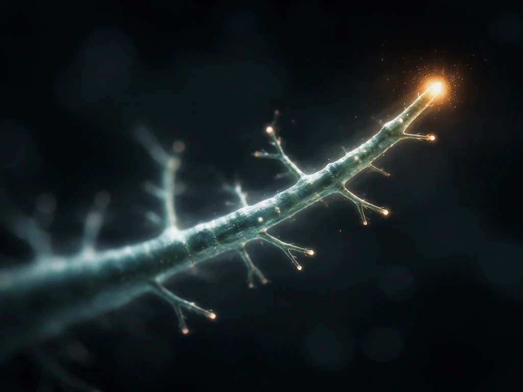
Dendrites grow by extending their tips outward, branching laterally from existing shafts, and stabilizing or retracting those branches depending on signals from the environment and from activity in the circuit. The whole process is driven by cytoskeletal remodeling inside the neuron and guided by molecular signals outside it. Growth is not unlimited: a mix of intrinsic programs, homeostatic feedback, and inhibitory cues in the extracellular space all act as brakes. Understanding those brakes is just as important as understanding what pushes growth forward.
How Do Dendrites Grow? Mechanisms and Practical Guide

Marcus Whitmore
14 May 2026
What dendrites are and where they actually grow
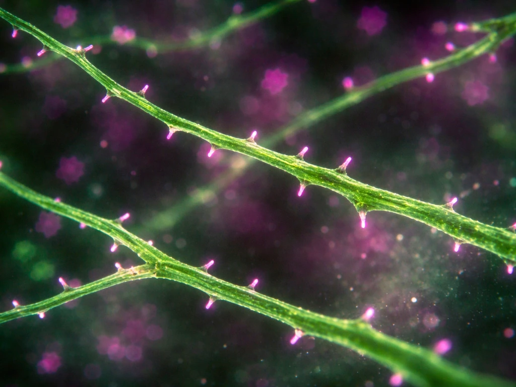
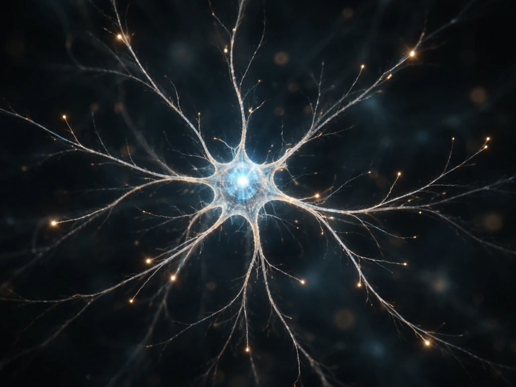
A neuron typically sends out one long axon that delivers signals to other cells, and several shorter dendrites that mainly receive input from incoming axon terminals. Think of the axon as the output cable and the dendrites as the antenna array. Dendrites are not just passive receivers though. They are dynamic structures that grow, branch, and form dendritic spines, which are the tiny protrusions where most excitatory synapses sit.
Most dendrite growth happens during early brain development, when neurons are differentiating and wiring up into circuits. But it does not stop there. In regions like the hippocampus, dendrites continue to remodel throughout life. Dendritic spines turn over continuously, with in vivo imaging showing ongoing spine gain and loss across weeks in adult mice. So 'where do dendrites grow?' has two honest answers: during development in nearly every brain region, and during plasticity in regions that remain structurally flexible into adulthood.
Do dendrites actually grow, and what limits them
Yes, dendrites grow, but not in a simple 'keep extending until told to stop' way. Even during peak development, dendrite tips lengthen, shorten, pause, and branch, all at the same time. In sensory neuron studies, the branching rate per unit of existing branch length drops roughly 10-fold between 24 and 96 hours of growth. That slowdown is not a failure. It is the system converging on a mature, stable arbor.
Several overlapping constraints prevent runaway growth. Transcription factors define which synaptic layers a neuron's dendrites can even reach. Morphological limits mean that only axons physically close enough to a dendrite can form synapses with it. Homeostatic plasticity mechanisms, including synaptic scaling, tune overall activity levels to prevent circuits from becoming overexcited or silent. If dendrites grew without limit, the circuit would destabilize. The limits are features, not bugs.
The cell biology: how a dendrite tip actually moves and branches
Dendrite extension is fundamentally a cytoskeleton problem. Two main structural systems do the work: actin filaments at growing tips and branch points, and microtubules running along the length of the dendrite. What makes dendrites structurally distinct from axons is that their microtubules have mixed polarity, with both plus and minus ends pointing toward the tip. That mixed polarity supports branching and allows organelles, including Golgi outposts, to be distributed throughout the arbor rather than just at the soma.
At the growing tip, the Arp2/3 complex (activated by the WAVE regulatory complex) nucleates new branched actin filaments. This creates the pushing force that moves the tip forward or initiates a lateral branch. Microtubule-associated proteins like MAP2 and MAP1A stabilize the microtubule tracks behind the growing front, locking in gains. Local protein synthesis, supported by ribosomes and Golgi outposts distributed in the dendrite, means the branch does not have to wait for every building block to be shipped from the soma. Growth is partly autonomous.
Rho-family GTPases act as molecular switches that translate external signals into cytoskeletal changes. Rac and Cdc42 generally push toward spine formation and branch stabilization. Rho tends to push toward retraction. The balance between these pathways at any given moment determines whether a dendritic tip advances, branches, or pulls back.
What tells a dendrite to grow, stop, or branch
Dendrite growth is steered by a combination of activity-dependent signals and molecular guidance cues in the extracellular space. These are not separate systems. They talk to each other constantly.
Signals that promote growth
- BDNF acting through TrkB receptors is one of the strongest pro-growth signals. Full-length TrkB increases dendritic branching. Truncated TrkB (the T1 isoform) triggers elongation of existing dendrites instead. The same ligand, different receptor isoform, different growth outcome.
- Calcium signaling links neural activity directly to structural change. When a neuron fires, calcium entering through NMDA receptors can activate transcription programs that drive dendritic outgrowth globally, and also stabilize local cytoskeleton at active branches.
- Semaphorin 3A, via the NRP1/PlexinA receptor complex and downstream Farp1, increases dendritic complexity. The mechanism involves remote intracellular signaling that reorganizes the actin cytoskeleton at branching points.
- Netrin-1 influences dendritic arbor remodeling in vivo beyond its classic role in axon guidance, with effects that depend on gradients and relative concentration changes rather than absolute levels.
- cAMP modulates TrkB trafficking and phosphorylation, which affects how well BDNF signaling actually reaches dendritic spines in mature neurons. Raising cAMP can overcome some inhibitory signals.
Signals that inhibit or limit growth
- Chondroitin sulfate proteoglycans (CSPGs) in the extracellular matrix form a molecular brake on structural plasticity. Perisynaptic CSPGs restrict dendritic spine motility, and their levels go up around CNS injury, which is part of why regeneration is so hard.
- Myelin-associated inhibitors, including Nogo, MAG, and OMGp, cause growth cone collapse and suppress neurite extension. PKC signaling mediates a significant part of their inhibitory effect by activating Rho.
- Transcription factor programs set hard boundaries. In Drosophila T4/T5 neurons, SoxN and Sox102F restrict dendrites and axons to single synaptic layers. Similar layer-specific constraints operate in vertebrate cortex.
- Homeostatic mechanisms detect when activity has climbed too high or dropped too low and scale synaptic strengths multiplicatively to push it back toward a set point. This acts as a structural brake on runaway growth driven by Hebbian plasticity.
How to actually encourage dendrite growth in experiments

If you are studying dendrite growth in a lab or classroom context, you are working with one of a few setups: dissociated primary neuron cultures, organotypic slice cultures, or in vivo imaging in an animal model. The principles are the same across all of them. You are trying to tip the balance of the growth-promoting vs. growth-limiting signals described above.
Optimizing the growth environment
- Coat your substrate. Poly-D-lysine (PDL) is standard for promoting neuron adhesion and maturation in culture. Laminin is often added alongside it for extra ECM support. Without a good substrate, neurons do not adhere well and dendritic processes fail to extend properly.
- Use BDNF. Adding exogenous BDNF to cultured hippocampal or cortical neurons consistently increases primary dendrite number and total dendritic complexity. The TrkB signaling axis (specifically the Shc pathway, not PLC-γ) is the key effector for BDNF-induced dendritic outgrowth.
- Manage your inhibitory environment. If you are testing pro-growth interventions against a realistic inhibitory background, you can add CSPGs or myelin extract to the culture medium to create a constrained substrate. If you want to maximize growth, minimizing extracellular CSPG levels (or using chondroitinase ABC to digest them) makes the substrate more permissive.
- Consider glial support. Glia are not just bystanders. Glial abundance affects how BDNF influences dendritic growth, and glia can release soluble cues that either support or inhibit dendritic arborization depending on neuron type. Mixed cultures with appropriate glia often show more mature dendritic morphology than pure neuron cultures.
- Drive activity carefully. Calcium-dependent signaling links network activity to dendritic growth, so chronically silencing neurons (with TTX, for example) or chronically overexciting them both disturb normal dendritic development. A physiological range of activity supports the best growth outcomes.
What to measure as evidence of growth

Sholl analysis is the go-to method. You draw concentric circles at increasing distances from the soma and count how many dendrite branches intersect each circle. From that profile you can extract total dendritic length, maximum reach from the soma (domain span), and branch distribution. For spines specifically, chronic two-photon imaging lets you track the same dendrite over days or weeks and calculate a turnover rate: the number of spines gained plus lost divided by twice the total spine count. A higher turnover rate means more structural remodeling is happening.
Why your experiment might not be working
Here are the most common failure modes when dendrite growth experiments produce underwhelming results, and what the biology tells you about each one.
| Problem | Likely biological reason | What to try |
|---|---|---|
| Little to no process extension | Poor substrate adhesion or an inhibitory ECM baseline | Switch to PDL + laminin coating; reduce serum or ECM inhibitors in the medium |
| BDNF added but no branching increase | TrkB may not be trafficked to spines without cAMP signaling support; truncated TrkB may dominate | Co-apply a cAMP elevator (e.g., forskolin); verify TrkB isoform expression in your cell type |
| Dendrites extend but retract quickly | Rho-pathway activation may be dominating Rac/Cdc42 activity, driving retraction | Check if myelin or CSPG contamination in medium is activating PKC/Rho; consider a Rho inhibitor in the assay |
| Growth stops early in culture | Normal developmental slowdown as branching rate per unit length drops over time; this is expected | Measure at multiple time points; compare to a branching-rate curve, not just an endpoint |
| High variability between wells | Glial density varies and strongly modulates BDNF responsiveness and dendritic outcomes | Standardize glia seeding density; use defined co-culture ratios |
| Activity-dependent manipulation had no effect | Calcium signaling pathway may be clamped by homeostatic mechanisms compensating for the perturbation | Extend the manipulation timeline; homeostatic responses operate on slower timescales than acute Hebbian changes |
One thing worth keeping in mind: a lot of 'failed' dendrite growth experiments are actually showing you that homeostatic brakes work exactly as they should. If you chronically stimulate neurons and dendrites do not keep growing without limit, that is the circuit protecting itself. The goal in studying this system is usually to understand which lever you are pulling, not to force unlimited growth that the cell will resist anyway.
Connecting the pieces
Dendrite growth is a conversation between the neuron's internal growth machinery (cytoskeleton, transcription factors, local translation) and external cues (BDNF, semaphorins, netrins, CSPGs, myelin inhibitors, and the activity of neighboring cells). Myelin-associated inhibitors of axonal regeneration include Nogo, MAG, and Omgp, which induce growth cone collapse and inhibit neurite outgrowth, with cyclic AMP described as able to overcome MAG inhibition myelin inhibitors. Neither side alone determines the outcome. That is why isolating single variables in experiments is both necessary and tricky. The same signal (semaphorin 3A, for example) that promotes dendritic complexity through one receptor pathway in one developmental context can have inhibitory effects in another. Context matters enormously.
If you want to dig deeper into the molecular triggers behind dendrite remodeling, the question of what specifically causes dendrites to grow covers the upstream signaling cascades in more detail. These upstream signaling cascades help explain what causes dendrites to grow in different brain regions and developmental stages.
And if you are thinking about dendrite growth in the context of how new synapses form, the mechanisms by which dendrites grow and new synapses are established are closely linked topics worth exploring together. For those interested in the broader question of how all brain cells develop and grow, not just their dendritic processes, the question of how brain cells grow overall puts dendritogenesis in a wider cellular context.
For those interested in the broader question of how all brain cells develop and grow, the question of how brain cells grow overall complements what this explains about dendritic remodeling.
FAQ
Do dendrites grow continuously, or only during development?
Dendrites remodel in two phases, early wiring during development and ongoing structural plasticity later. Even in adult tissue, spine gain and loss can continue, but the overall arbor often shifts toward a more stable shape with lower branching turnover.
Why might I see dendrite “growth” in culture but not in an animal model?
The extracellular brake strength and activity patterns differ across systems. Slice and in vivo environments include additional guidance sources and inhibition, so the same molecular cue can produce weaker extension or more retraction unless the circuit activity and cell maturity match the experimental window.
What’s the most common reason dendrites stop advancing even when I add a growth-promoting factor?
Homeostatic counterbalancing. If you chronically raise activity or excitatory drive, synaptic scaling and other feedback can shift the balance toward stabilization or retraction, so dendrite tips may pause or branch less despite the factor being present.
How can I tell whether my treatment is changing dendrite length versus branching?
Use a readout that separates effects. Sholl analysis gives branch intersections versus distance, but pairing it with total length measurements and, when possible, spine density or spine turnover helps distinguish “more branches,” from “longer reach,” from “same reach but more spines.”
Are dendrites actually guided by the same signals that guide axons?
They share some broad molecular themes, but the decision logic is different. Dendrites integrate local guidance cues with ongoing network activity, and context can flip a cue’s effect from promoting to inhibiting depending on receptor pathway engagement and developmental stage.
Why do dendritic tips sometimes retract instead of extending further?
Rho-family signaling balance can tilt toward retraction, and stabilization signals may not persist long enough to lock in gains. Tip behavior can include advance, pause, and pullback simultaneously across different segments, so averaged outcomes can hide local retraction events.
What controls whether new branches become stable spines versus retracting quickly?
Branch stability depends on whether an appropriate synaptic partner and synaptogenic conditions are met, not just actin-driven outgrowth. Spine formation often requires coordinated signaling that couples extracellular cues to internal cytoskeletal programs, so a branch can extend but fail to remain if synaptic matching is limited.
How important is local protein synthesis for dendrite growth in experiments?
It can be decisive for local autonomy. If you disrupt synthesis or trafficking, tips may struggle to stabilize and remodel even if initial extension occurs, leading to growth that looks transient or fails to convert into durable branch architecture.
What should I watch out for when interpreting “increased Sholl intersections”?
More intersections can reflect increased branching, but it can also come from a redistribution of branches at specific radii. Always check maximum reach and branch distribution together, and confirm with morphology metrics like total length or spine density to avoid overinterpreting a single Sholl feature.
If spines are turning over faster, does that mean dendrites are growing more?
Not necessarily. Spine turnover can be high even when dendritic reach is stable, reflecting synaptic remodeling rather than arbor expansion. Separating spine dynamics from dendritic domain span helps you attribute changes to synapses versus structural skeleton remodeling.
Next Article
Dendrites Grow and New Synapses Are Formed: Mechanisms
How dendrites grow and new synapses form: activity, trophic signals, adhesion, cytoskeleton changes, and how to measure