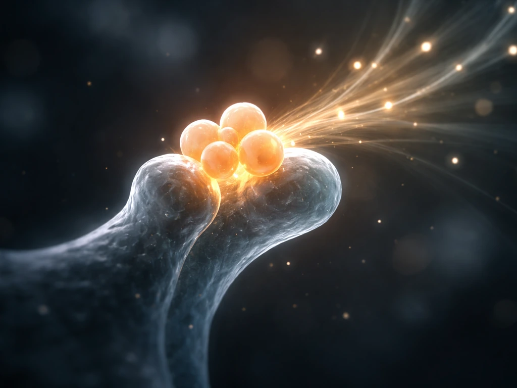
Brain cells grow in two distinct ways: they multiply (new cells are made through cell division) and they expand (existing cells grow bigger, sprout branches, and wire up connections). Both happen constantly during fetal development and early childhood, slow down dramatically after that, but never fully stop. Even in adulthood, your brain keeps reshaping itself through a process called plasticity, and a small but real trickle of new neurons keeps forming in at least one region of your hippocampus. Understanding which kind of "growth" you're asking about changes everything about what's actually possible.
How Do Brain Cells Grow From Birth to Adulthood

Marcus Whitmore
4 May 2026
What "brain cell growth" actually means
"Brain cells" covers a lot of ground. There are neurons, the cells that send electrical signals, and there are glial cells (including astrocytes, oligodendrocytes, and microglia), which support, insulate, and protect neurons. Both types grow, but in very different ways and on very different timelines.
When most people ask how brain cells grow, they're usually mixing together three separate questions: How do new brain cells get made? How do individual cells get bigger and more complex? And can the brain repair itself after injury? These aren't the same question, and they don't have the same answer.
- Neurogenesis: the production of brand-new neurons from stem or progenitor cells
- Gliogenesis: the production of new glial cells (happens more readily than neurogenesis in adults)
- Neurite outgrowth: individual neurons growing longer axons and dendrites to reach targets
- Synaptogenesis: neurons forming new connection points (synapses) with each other
- Regeneration: regrowing damaged or severed axons after injury (extremely limited in the adult brain)
Most of the exciting plasticity you hear about in adults falls into the synaptogenesis and neurite outgrowth categories, not the "making brand-new neurons" category. That distinction matters a lot when you're evaluating claims about brain-boosting habits.
How the brain builds itself: neurogenesis and the cell cycle
Early in fetal development, the entire nervous system is built from a thin sheet of cells called the neuroepithelium. These cells divide rapidly through mitosis, but the way they divide is carefully controlled to produce the right mix of neurons and glia in the right places at the right times.
There are two main division strategies at play. In direct neurogenesis, a neural stem cell divides and one daughter cell immediately becomes a neuron. In indirect neurogenesis, the stem cell first produces a transient-amplifying progenitor, which divides a few more times before the resulting cells differentiate into neurons. Indirect neurogenesis lets the brain scale up cell numbers fast, like compound interest on a savings account. The outcome depends on factors like cell-cycle timing, how chromosomes and cell fate molecules are distributed between daughter cells (called asymmetric inheritance), and a process called interkinetic nuclear migration, where the nucleus physically moves up and down inside the cell as it divides.
At some point during development, the same progenitor cells switch from making neurons to making glial cells. This neurogenesis-to-gliogenesis transition is driven by signaling pathways, especially JAK-STAT and Notch. Notch is particularly important: when a neighboring progenitor sends a Notch signal, the receiving cell ramps up Hes and Hey proteins, which dial down "become a neuron" genes and keep the cell in a progenitor or glial fate. Disrupt this system and you get premature differentiation and disorganized brain structures.
Three other signaling systems coordinate the whole process. Sonic Hedgehog (Shh) controls proliferation and patterning, especially in the ventral brain. Wnt signaling influences transcription, cell cycle control, and asymmetric division. BMP signaling helps define dorsal regions and cross-talks with both Wnt and Shh to keep everything in balance. Think of them as the architectural team deciding not just how many cells to build, but where each type goes.
How neurons and glia actually get bigger
Once a neuron is born, its work is just starting. A newborn neuron is a small, round cell. Over weeks to months it grows dramatically: the cell body (soma) enlarges, and it sprouts long processes called neurites, which eventually differentiate into a single long axon and multiple branching dendrites. This process is called neurite outgrowth, and it's one of the most visually dramatic forms of growth in all of biology.
Axons are guided to their targets by a combination of attractive and repulsive molecular signals in the environment, including proteins like semaphorins, ephrins, and netrins. The tip of a growing axon, called the growth cone, samples these signals constantly and steers accordingly, like a ship following a magnetic compass through fog. When the axon reaches the right target, it forms a synapse, a junction where chemical signals can pass from one neuron to the next.
Dendrites follow a different growth program. In simple terms, dendrites grow by branching and remodeling their structure in response to signaling and activity. They branch and elaborate over time, partly driven by activity. That activity-dependent remodeling is how dendrites grow and new synapses are formed in the circuits that matter for learning. The more a neuron is stimulated, the more its dendrites tend to branch and form new spines, which are the small protrusions that house individual synapses. This is the structural basis of learning: when you practice something, the relevant neurons are literally reshaping themselves to make that pattern of activity easier to repeat.
Glial cells grow differently again. Oligodendrocytes wrap myelin (a fatty insulating sheath) around axons, and the process of myelination continues well into a person's twenties. Astrocytes extend long processes that contact synapses and blood vessels, helping regulate the chemical environment around neurons. Both types respond to activity and injury by changing their shape and gene expression.
The signals that tell brain cells when and how to grow
Growth in the brain is never random. It's tightly controlled by signals from outside the cell (extrinsic signals) and from the cell's own genes (intrinsic programs). These two layers interact constantly.
Molecular pathways and hormones
Brain-derived neurotrophic factor (BDNF) and its receptor TrkB are arguably the most important growth regulators in the mature brain. BDNF supports the survival, growth, and differentiation of neurons, promotes synapse formation, and drives long-term plasticity through a downstream pathway involving mTOR, which coordinates protein synthesis in dendrites. When local translation is triggered at a synapse, the dendritic spine can change its shape and strength within minutes, without waiting for instructions from the cell body. BDNF-TrkB signaling underpins virtually every experience-dependent change in brain structure.
Stress hormones push in the opposite direction. Chronic exposure to glucocorticoids (the cortisol family) shrinks dendrites in the hippocampus, reduces dendritic branching in CA1 and dentate gyrus neurons, and suppresses adult neurogenesis. It's not a subtle effect. Prolonged stress measurably alters the physical structure of hippocampal neurons, which helps explain why chronic stress impairs memory.
Activity-dependent remodeling
One of the most important things to understand about brain cell growth is that activity itself is a growth signal. When neurons fire together repeatedly, the synapses connecting them are strengthened, new spines form, and in some cases axons sprout small branches (called collateral sprouting or reactive synaptogenesis) to reinforce a circuit. This is why learning and practice genuinely change brain structure, not just function. The extracellular matrix around neurons also plays a role: structures called perineuronal nets (PNNs) wrap around certain neurons and regulate how plastic they can be. In animal models, enzymatically digesting PNNs increases plasticity and can affect how readily new memories form and interfere with old ones.
What stops brain cells from growing indefinitely
If BDNF promotes growth and activity reshapes connections, why doesn't the brain just keep growing without limit? Several mechanisms act as brakes.
First, the physical environment imposes limits. The brain sits inside a rigid skull, so volume is capped. Caloric and oxygen availability constrain how metabolically active new growth can be. The brain already consumes about 20% of your body's resting energy despite being roughly 2% of body mass, so adding more cells or more connections has a real metabolic cost.
Second, the molecular environment of the adult brain actively suppresses regrowth, especially after injury. Two major classes of inhibitors are at work: myelin-associated inhibitors (including proteins called Nogo, MAG, and OMgp) and chondroitin sulfate proteoglycans (CSPGs) produced by the glial scar that forms around an injury site. These molecules bind receptors on axons and activate the RhoA/ROCK signaling pathway, which essentially tells the growth cone to stop and retract. In preclinical models, blocking these inhibitors (using neutralizing antibodies, ROCK inhibitors, or enzymes like chondroitinase ABC that break down CSPGs) can improve axon regrowth, but translating this to human clinical use is still an active research challenge.
Third, mature neurons have reduced intrinsic growth capacity compared to developing ones. A fetal neuron is primed to grow long axons. An adult neuron has largely switched off those developmental programs. So even if you clear the external inhibitors, the neuron itself may not respond as vigorously as it would have during development. Both the cell's internal state and its external environment need to be permissive for regrowth to occur.
Can you actually grow new brain cells after childhood?
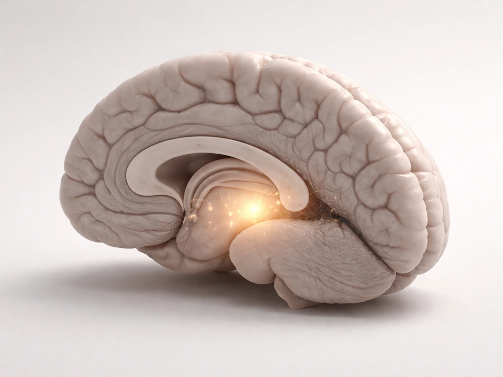
Yes, but with important caveats. Adult neurogenesis in humans is real but geographically limited. It occurs most robustly in one region: the dentate gyrus of the hippocampus, which is involved in memory formation and spatial navigation. There is also evidence for neurogenesis in the subventricular zone (SVZ), which lines the ventricles. Estimates from human studies suggest roughly 700 new neurons are added to the hippocampus per day in adults, corresponding to a turnover rate of about 1. This estimate of human adult hippocampal neurogenesis, about 700 new neurons per day per hippocampus, is supported by research on the dynamics of adult human hippocampal neurogenesis suggggest roughly 700 new neurons are added to the hippocampus per day in adults. 75% per year in the renewing population. That's not nothing, but it's also not a flood of new cells replacing damaged tissue.
These new neurons start out electrically different from mature neurons: they receive GABAergic inhibitory input that's actually excitatory early in their development, before they integrate fully into the existing circuit. It takes weeks to months for a new neuron to mature enough to participate meaningfully in memory function. So even when new neurons form, their contribution is slow and gradual.
Outside the hippocampus, the picture is much more restricted. Most of the cerebral cortex, which handles reasoning, language, and perception, does not produce new neurons in adulthood. Damage there is met with glial scar formation, some sprouting of surviving axons, and synaptic remodeling, but not true neuronal replacement. This is why stroke or traumatic brain injury can cause permanent deficits even though the brain does mount a recovery response.
It's also worth noting that glia, especially oligodendrocytes and astrocytes, are produced more readily than neurons throughout adulthood. When the brain is injured, gliogenesis ramps up as part of the repair response, even when neurogenesis doesn't.
A quick comparison: neuron growth vs glial growth vs axon regrowth
| Type of growth | When it's most active | Happens in adult humans? | Main limiting factors |
|---|---|---|---|
| New neuron production (neurogenesis) | Fetal development and early childhood | Yes, but limited to hippocampus/SVZ (~700 neurons/day in dentate gyrus) | Loss of progenitor populations, inhibitory environment, reduced intrinsic capacity |
| New glial cell production (gliogenesis) | Fetal through adolescence; reactive after injury | Yes, more readily than neurogenesis | Less restricted; astrocytes and oligodendrocytes produced throughout life |
| Axon and dendrite growth (neurite outgrowth) | Fetal through early postnatal | Yes, partial (sprouting, spine formation, synaptogenesis) | Perineuronal nets, myelin inhibitors, CSPGs, metabolic cost |
| Axon regrowth after injury | Very limited in adult CNS | Extremely limited; better in peripheral nervous system | Nogo/MAG/OMgp inhibitors, glial scar (CSPGs), reduced intrinsic growth programs |
What you can actually do today to support brain cell health
You can't regrow a damaged cortex, but you have more control over brain plasticity than most people realize. The following factors have the strongest evidence behind them for supporting neurogenesis, synaptic remodeling, and overall brain cell health in adults.
Aerobic exercise
This is the single most replicated finding in the adult neurogenesis literature. Aerobic exercise reliably increases BDNF expression in the hippocampus, and higher BDNF levels support all the steps of new neuron development: proliferation, differentiation, maturation, and integration into existing circuits. Running, cycling, swimming, or any sustained cardio activity done consistently (most days per week) is your most direct lever for supporting the cellular growth side of brain health.
Sleep
Sleep is when a huge amount of synaptic consolidation happens. During sleep, the brain prunes weaker synapses and strengthens important ones, a process tied to the synaptic homeostasis hypothesis. Sleep deprivation measurably alters dendritic spine density in animal models and disrupts BDNF expression. Even short-term sleep loss changes the molecular markers of plasticity. Prioritizing 7 to 9 hours of quality sleep isn't just about feeling rested; it's when your synaptic remodeling actually runs.
Managing chronic stress
Chronic stress is genuinely bad for brain cell growth, not in a metaphorical way but in a measurable, structural way. Sustained glucocorticoid exposure shrinks hippocampal dendrites and suppresses neurogenesis. That's not permanent damage in most cases, but it does mean that reducing chronic stress (through whatever sustainable means works for you) directly supports the conditions that allow brain cells to grow and maintain healthy structure.
Novelty, learning, and cognitive challenge
Activity-dependent plasticity is real. Learning a new skill, navigating unfamiliar environments, practicing a musical instrument, or acquiring a second language all drive dendritic spine formation and synaptogenesis in the relevant circuits. The brain responds to demand by restructuring itself to meet it. Doing the same tasks on autopilot doesn't provide the same stimulus. New and challenging experiences drive more remodeling.
Nutrition basics
The brain is metabolically expensive and requires reliable glucose, oxygen, and key micronutrients. Omega-3 fatty acids (especially DHA) are structural components of neuronal membranes. B vitamins support myelination and neurotransmitter synthesis. Severe caloric restriction or nutrient deficiencies impair neurogenesis and synaptic function. There's no "brain growth superfood," but a diet that supplies adequate calories, healthy fats, and micronutrients removes metabolic obstacles to normal plasticity.
What to avoid
- Chronic alcohol use: suppresses hippocampal neurogenesis and damages myelin
- Chronic sleep deprivation: disrupts synaptic remodeling and BDNF expression
- Prolonged social isolation: associated with reduced plasticity and elevated stress hormones in animal models
- Sedentary lifestyle: removes the primary trigger (aerobic exercise) for BDNF-driven neurogenesis
- Unmanaged chronic stress: sustained glucocorticoid exposure directly shrinks hippocampal dendrites
When to seek medical guidance
If you're asking about brain cell growth in the context of a brain injury, stroke, neurodevelopmental concern, or a condition like epilepsy or a brain tumor, lifestyle habits are not a substitute for medical evaluation and treatment. The inhibitory mechanisms around injury (glial scar, myelin inhibitors) are beyond what any habit-based intervention can currently overcome. Neurologists and rehabilitation specialists can guide evidence-based recovery strategies that go beyond what's covered here. Early intervention after brain injury consistently produces better outcomes than delayed treatment, so don't wait.
For deeper dives into specific parts of this story, the mechanics of how dendrites specifically grow and branch, what triggers new synapse formation, and what conditions cause dendrites to change shape are all rich topics in their own right, each with its own set of molecular players and practical implications for learning and memory. If your goal is to actually grow neurons in a lab, you’d be looking at stem cell and culture techniques rather than relying on adult brain plasticity.
FAQ
When people say “brain cells grow” from habits, are they always talking about making new neurons?
Not usually. Most lifestyle effects are about strengthening and remodeling existing connections, like synapse formation and dendritic spine changes. New neuron generation in adults is limited mainly to the hippocampal dentate gyrus and develops slowly, so improvements from habits typically reflect circuit plasticity more than neuron replacement.
How long does it take for new adult-born neurons to meaningfully affect memory?
Even after new neurons are born in the hippocampus, they need weeks to months to mature enough to integrate into existing networks. During that window, they are often not contributing strongly to memory performance, so you should not expect rapid, day-to-day changes from adult neurogenesis alone.
Is it possible to “force” the adult cortex to regrow neurons after injury?
Current evidence does not support reliably replacing neurons across most of the cortex in adulthood. After injury, the environment tends to become inhibitory (for example, scar-related molecules and myelin inhibitors), so recovery relies more on surviving-circuit remodeling, axonal sprouting, and synaptic changes than true neuronal replacement.
What’s the difference between dendrite growth and synapse growth, and which one does learning change most?
Dendrite growth involves branching and remodeling of dendritic structure, while synapse growth reflects the formation and strengthening of synaptic connections, often visible as changes in spine number and spine strength. Learning mainly drives activity-dependent synaptic and spine changes in relevant circuits, with dendritic remodeling as part of the same structural adaptation process.
Do stress and sleep affect the whole brain equally, or are there regional differences?
They are not equal across regions. Chronic glucocorticoids have especially strong documented effects on hippocampal dendrites and can suppress adult neurogenesis there, while the cortex often shows different limiting factors after injury. Sleep disruption can affect plasticity markers broadly, but its impact can still be stronger in networks critical for memory consolidation.
How can I tell whether an intervention is improving plasticity versus just boosting motivation or attention?
Look for changes that track with learning and consolidation rather than short-term alertness. For example, skills that improve with practice over weeks, or memory that benefits from adequate sleep, suggest circuit remodeling. Also consider whether performance gains persist after a rest period, since consolidation depends on synaptic strengthening processes.
Can too much exercise or too much training backfire for brain plasticity?
Yes, especially if it causes chronic sleep loss, persistent overtraining stress, or inadequate recovery. Since stress hormones and poor sleep can reduce growth-promoting conditions (like hippocampal structural support), the “dose” matters, consistency matters, and recovery is part of supporting plasticity rather than ignoring it.
What role do diet and micronutrients play if there is no “brain growth superfood”?
Diet mainly removes constraints. Adequate calories and key building blocks support normal synapse formation and neuronal metabolism. If you are under-eating, deficient in important micronutrients, or chronically metabolically stressed, the brain may not have sufficient resources to sustain healthy remodeling, even if your activity level is high.
Does “learning new skills” always increase brain growth signals, or does it depend on how you practice?
It depends on engaging the relevant circuits with challenge and attention. Repeating a task on autopilot provides weaker training signals than practicing with novelty, feedback, or increasing difficulty. The brain responds most strongly when neural activity repeatedly co-occurs in patterns the circuit needs to update.
Are there medical situations where lifestyle changes can’t overcome the biology of regrowth?
Yes. After major injury, stroke, tumors, or neurological conditions, inhibitory environments and disease-specific mechanisms often require medical care and rehabilitation strategies. Lifestyle can support general brain health and recovery capacity, but it cannot substitute for targeted treatment, especially when regrowth is limited by scar and myelin-associated inhibitors.
Next Article
What Controls When and How Fast Cells Grow and Divide
Learn what controls cell division timing and speed: checkpoints, signals, resources, DNA damage, stress, and failure lea