Mature red blood cells do not grow, and they cannot divide or reproduce. Once a red blood cell reaches full maturity, it has already shed its nucleus and most of its internal machinery, leaving it with no way to copy DNA, run cell division, or even repair itself. What the body does instead is constantly manufacture brand-new red blood cells in the bone marrow to replace the ones that wear out after about 120 days. So when people ask whether red blood cells grow, the honest answer is: the precursor cells that become RBCs absolutely go through growth and division, but the finished product does not.
Do Red Blood Cells Grow? How RBCs Form and Mature

Marcus Whitmore
22 May 2026
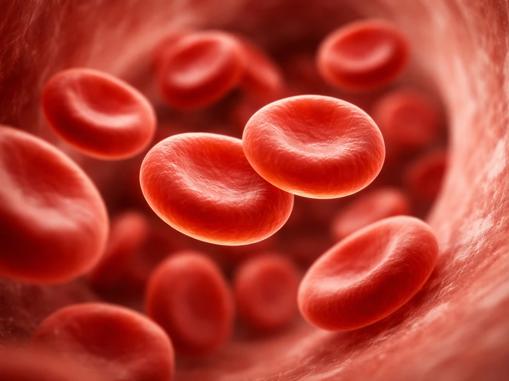
What red blood cells actually are (and why "growing" doesn't apply)
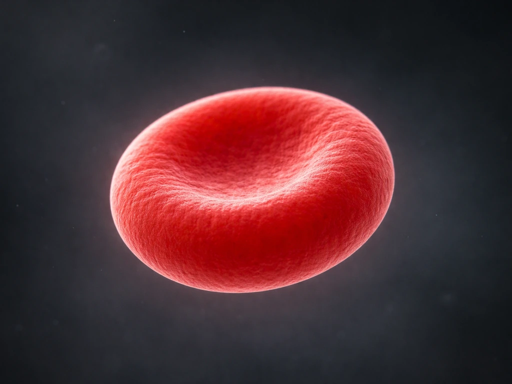
A mature red blood cell is basically a stripped-down delivery vehicle. It has a distinctive biconcave disc shape, it carries hemoglobin, and it is built for one job: shuttling oxygen from your lungs to your tissues and hauling carbon dioxide back. To do that job efficiently, the cell goes through a radical transformation during development. It ejects its nucleus, dismantles its mitochondria, gets rid of its endoplasmic reticulum and Golgi apparatus, and sheds essentially every organelle that nucleated cells rely on to grow, divide, and maintain themselves.
That stripping-down is not a flaw. It frees up more internal space for hemoglobin and makes the cell flexible enough to squeeze through the narrowest capillaries. But it means the mature RBC is, in a meaningful biological sense, a cell that has voluntarily dismantled its own growth machinery. Without a nucleus there is no DNA. Without DNA there is no RNA. Without RNA there is no protein synthesis and no pathway to mitosis. The cell literally cannot grow even if the conditions were ideal for it.
This puts RBCs in a very different category from most cells you might think about. Skin cells divide and renew. Liver cells can regenerate. Even muscle cells, which are largely post-mitotic, retain their nuclei. Red blood cells are one of the few cell types that genuinely have no internal toolkit left for any kind of growth after maturity.
How new red blood cells are made: erythropoiesis from start to finish
The process of making red blood cells is called erythropoiesis, and it happens almost entirely in the red bone marrow. Think of the marrow as a continuous manufacturing line that runs every single day of your life. At the start of the line are hematopoietic stem cells, which are multipotent cells that can commit to becoming any blood cell type. When they commit to the red blood cell path, they move through a series of increasingly specialized stages.
The hormone that kicks it all off: erythropoietin
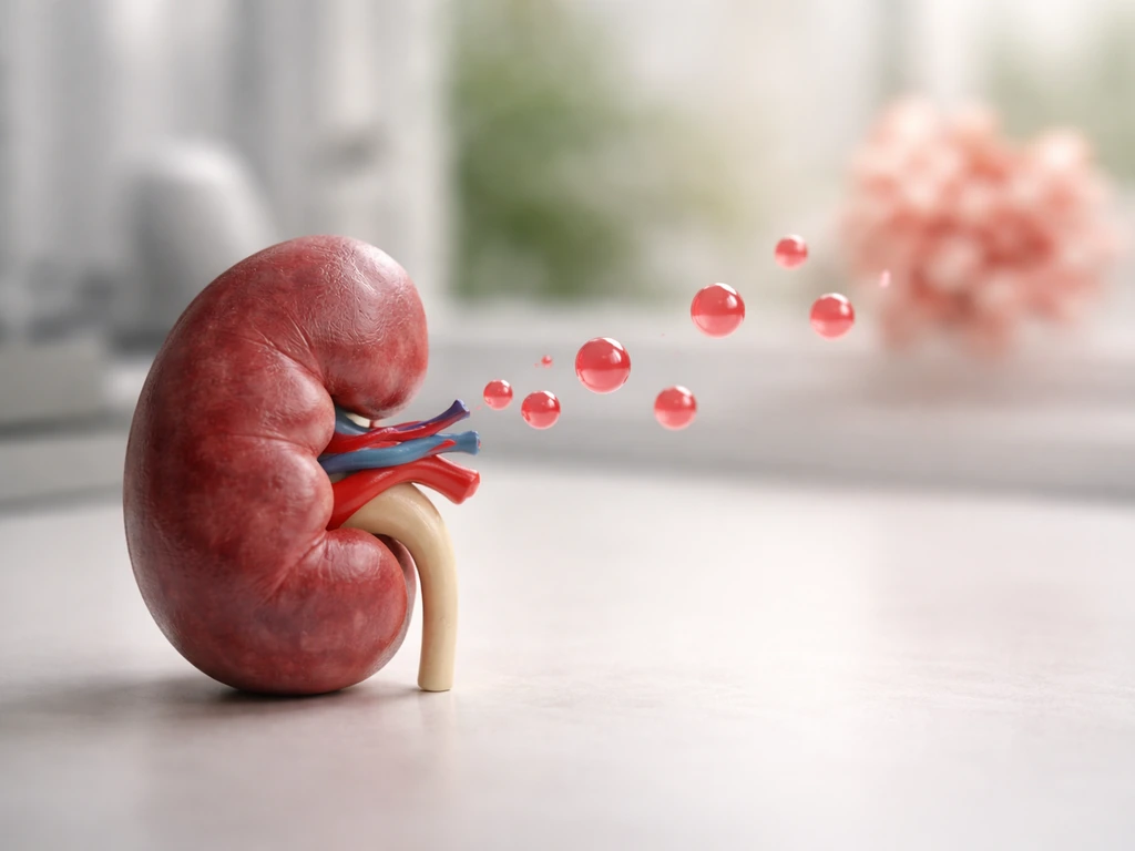
The signal driving this whole process is erythropoietin, almost always called EPO. When your blood oxygen drops, specialized fibroblast-like cells in the kidney detect the low oxygen levels and ramp up EPO production. The trigger is a protein called hypoxia-inducible factor 2 (HIF-2), which acts as a transcriptional switch: low oxygen activates HIF-2, HIF-2 cranks up EPO gene expression, and EPO floods the bloodstream and reaches the bone marrow. There, it tells erythroid progenitor cells to proliferate faster and mature more quickly. When oxygen levels recover, the signal dies down. It is a clean feedback loop that matches RBC production to the body's actual oxygen demand.
Cell division during development: the mitotic phase
Here is where the growth and division that people might associate with RBCs actually happens. The earliest recognizable erythroid cell, called the pro-erythroblast, undergoes roughly four to five rounds of mitosis. Each division produces daughter cells that are progressively smaller and more specialized: basophilic erythroblasts, then polychromatophilic erythroblasts, and finally orthochromatic erythroblasts. By the orthochromatic stage, the cell cycle has shut down completely. The cell arrests, its chromatin compacts, and it prepares for the final step: ejecting its nucleus.
That ejection event, called enucleation, produces two structures. One is the pyrenocyte, which is just the expelled nucleus wrapped in a thin membrane. The other is the reticulocyte, an anucleate young cell that still contains traces of RNA and a few residual organelles. The reticulocyte enters circulation and within about one day finishes maturing into the fully stripped-down red blood cell. It loses its remaining RNA and becomes the smooth, biconcave RBC you see in textbook diagrams. So any cell division associated with RBCs happens before enucleation, deep in the marrow, not in circulation.
How long RBCs last and what happens when they wear out
A mature red blood cell circulates for about 120 days. Over that time it takes a beating, literally. It gets squeezed through capillaries smaller than its own diameter, it cycles through changes in pH and oxygen pressure thousands of times, and its membrane gradually stiffens and oxidizes. Without a nucleus or ribosomes, it cannot make new proteins to repair damage. The wear accumulates.
Around the 120-day mark, aging RBCs start displaying molecular signals on their surface, including changes to a membrane protein called Band 3 and the attachment of complement proteins. These act as "eat me" flags that macrophages in the spleen, liver, and bone marrow recognize. The macrophages engulf the old cells, break them down, and recycle the iron and other components. This clearance is not a failure of growth; it is a programmed, orderly retirement system.
The key point is that nothing about this lifespan involves the RBC getting bigger, stronger, or more capable as it ages. There is no growth phase in maturity. The cell is effectively static from the moment it finishes maturing until the moment it is cleared.
More RBCs vs. bigger RBCs: it's always about production rate
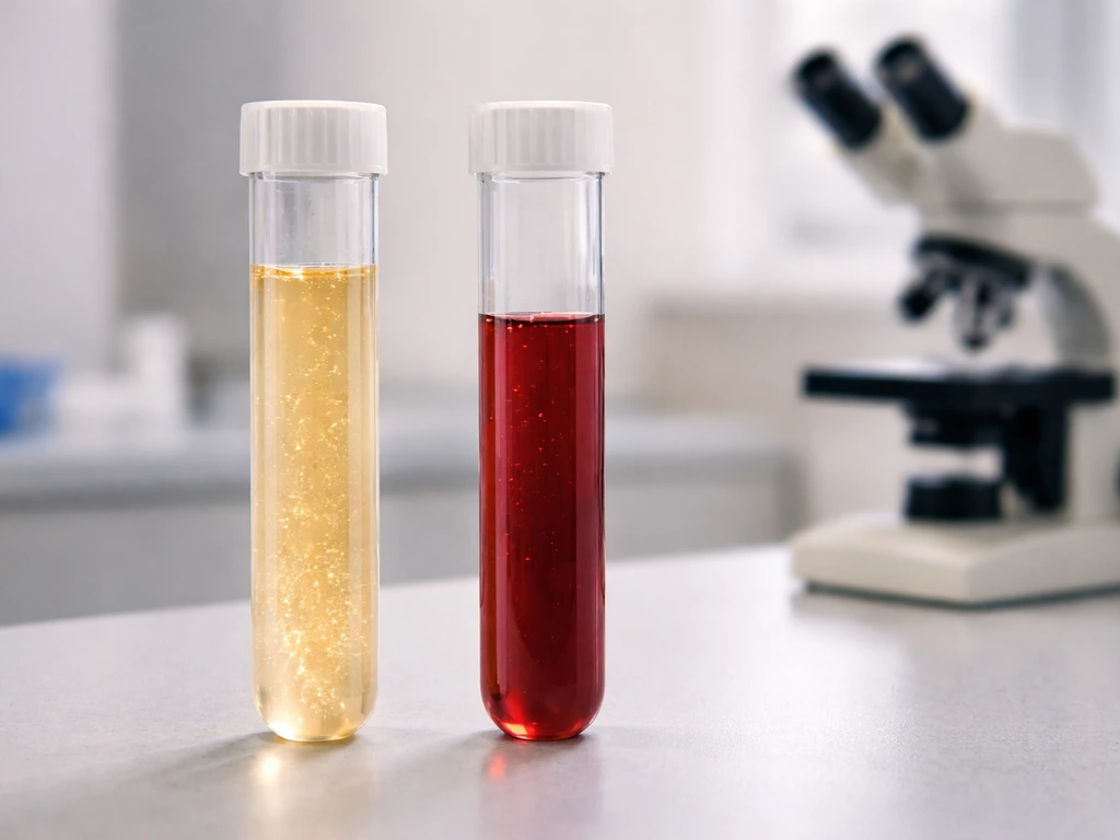
When the body needs more oxygen-carrying capacity, it does not grow existing red blood cells larger. It makes more of them. Healthy adults renew roughly 170 billion red blood cells every single day. That staggering number reflects a steady-state balance: about the same number of new reticulocytes are released from the marrow as old RBCs are cleared by macrophages.
You can track this production rate by measuring reticulocytes. Normally, reticulocytes make up about 1-2% of circulating red blood cells. If the percentage jumps, it means the marrow is accelerating production, usually in response to anemia, blood loss, or hemolysis. If the reticulocyte count stays low when RBC numbers are falling, it tells clinicians the marrow is not responding adequately, which points to conditions like iron deficiency, B12 deficiency, or bone marrow suppression. Importantly, the increased reticulocyte count appears about 3 to 4 days after an acute drop in RBC mass, reflecting the time it takes for the marrow to ramp up and release new cells, not for existing ones to change.
Anemia, in this light, is almost never a story about RBCs failing to grow after maturity. It is a story about production not keeping pace with destruction or loss. The treatment logic follows from that: fix the production problem (supply iron, B12, or folate; treat the underlying marrow issue; use EPO-stimulating agents in certain kidney disease cases) and the new RBC supply recovers.
Growing RBCs in a lab: what that actually means
Researchers and biotech companies do talk about "growing" red blood cells, which can sound confusing given everything above. What they mean is culturing the precursor cells, not the mature RBCs themselves. The goal is to start with hematopoietic stem cells or induced pluripotent stem cells (iPSCs) and push them through the same erythropoiesis process in a dish, ultimately producing enucleated red blood cells that could theoretically be used for transfusions.
One established approach uses a two-step liquid culture method. In the first phase, progenitor cells expand without EPO. In the second phase, EPO is added to drive the cells through differentiation toward orthochromatic normoblasts, followed by enucleation. Researchers have also developed systems that let them watch nascent reticulocytes finish maturing in culture, giving a window into that final transition from reticulocyte to fully mature RBC.
But this field still has real bottlenecks. Getting cells to complete enucleation efficiently, achieving the proper biconcave disc shape, avoiding immature RBC characteristics, and scaling production to clinically useful quantities are all ongoing challenges as of 2026. The point is that even in the lab, scientists are not making mature RBCs grow or divide. They are recapitulating the natural development process, just outside the body.
Why mature RBCs can't just keep dividing: the physics and biology of limits
It is worth stepping back and asking why terminal differentiation exists at all. Why would a cell permanently shut off its ability to divide? The answer involves both function and safety.
On the functional side, the same organelles that enable division take up space and resources that, in an RBC, are better spent on hemoglobin. A cell that kept its nucleus would be less efficient at its core job. On the safety side, cells that divide without restraint are the definition of cancer. Terminally differentiated cells like mature RBCs, neurons, and cardiac muscle cells exit the cell cycle permanently, which removes them from the population of cells that could accumulate mutations and proliferate uncontrollably. The cell-cycle machinery itself is effectively dismantled in these states, meaning growth signals from outside the cell have nothing to act on even if they arrive.
For RBCs specifically, the transcriptional programs driven by factors like GATA-1 and related epigenetic regulators orchestrate the shift from proliferation to maturation. Once those programs commit the cell to terminal differentiation, the process is irreversible. No external signal, no matter how strong, can push a mature red blood cell back into division. This is a fundamentally different situation from cells that simply happen to be resting and could in principle re-enter the cycle under the right conditions.
This kind of hard limit on cell division is a theme that runs across biology. If you are curious how other cell types navigate the tension between growth and restraint, it is worth comparing RBCs to skin cells or animal cells more broadly, where the growth rules are quite different because the nucleus is still present and the cell cycle is still available.
The bottom line: what "growing" means for red blood cells
| Stage | Does it grow or divide? | Key reason |
|---|---|---|
| Hematopoietic stem cell | Yes, proliferates actively | Nucleus intact, full cell-cycle machinery available |
| Pro-erythroblast to orthochromatic erythroblast | Yes, undergoes ~4-5 mitotic divisions | Still nucleated, driven by EPO signaling |
| Orthochromatic erythroblast (late) | No, cell cycle arrests | Terminal differentiation program activates, prepares for enucleation |
| Reticulocyte | No, finishes maturing only | Nucleus ejected, residual RNA cleared over ~1 day |
| Mature red blood cell | No growth or division | No nucleus, no DNA, no organelles for division or repair |
Red blood cells grow and divide only during their development in the bone marrow, while they still have a nucleus. In the case of red blood cells, that growth happens during development in the bone marrow, where cells divide and mature over days how fast do cells grow. Once enucleation happens, all growth stops permanently. The body compensates by running erythropoiesis continuously, replacing roughly 170 billion cells daily. If your RBC count is low, the answer is always found upstream in production, never in trying to make mature RBCs grow. That distinction matters whether you are studying for an exam, trying to understand a blood test result, or just curious about how one of your body's most abundant cells actually works. If you meant “growth” in the sense of resizing or expanding text within an Excel cell, you can adjust font size, enable wrap text, and format row height to fit the content make cells grow with text in excel.
FAQ
If mature red blood cells cannot grow, why do some labs talk about “reticulocyte response” or “recovery” after anemia?
Recovery usually means the marrow ramped up production and released more reticulocytes, not that older mature RBCs changed size or function. Reticulocytes rise first, then mature into RBCs over about a day, so the visible improvement lags behind the trigger by several days.
Can mature red blood cells repair themselves or replace damaged parts while they are circulating?
They cannot meaningfully repair themselves because they have no nucleus and no ribosomes, so they cannot synthesize new proteins needed for most repair processes. Damage accumulates until they are cleared by macrophages based on aging markers.
Why does the reticulocyte count rise 3 to 4 days after an acute RBC drop?
There is a delay between the initial loss and when newly produced cells reach circulation. After sensing the deficit, the marrow accelerates erythropoiesis, and the new cells need time to divide, differentiate, enucleate, and then mature from reticulocytes into RBCs.
If the body makes about 170 billion RBCs per day, does that mean daily production always stays the same?
No. Production changes with oxygen demand and RBC loss. Erythropoietin levels increase when oxygen delivery is low or when RBCs are being destroyed or lost, driving higher proliferation in the marrow; production drops when the trigger resolves.
Do higher hemoglobin levels always mean RBCs are “growing,” or can it be something else?
Higher hemoglobin can reflect increased production (often with a higher reticulocyte percentage), decreased destruction, or less blood loss. It is also possible for hemoglobin to look higher due to changes in plasma volume, so interpretation depends on the full blood count and the reticulocyte trend.
What happens in kidney disease where EPO signaling may be altered, does that change whether RBCs can grow?
It mainly affects production rate upstream, not the ability of mature RBCs to grow. Reduced EPO can lead to insufficient marrow output, so the reticulocyte response will be low even though existing RBCs cannot divide anyway.
Can you “grow” red blood cells outside the body for transfusions, and if so, what step is the real challenge?
Researchers can culture precursor cells through erythropoiesis, but the bottleneck is reliably completing enucleation and producing a sufficient number of fully mature, properly shaped enucleated RBCs. Immature characteristics or incomplete enucleation can limit usable output.
In blood tests, does a low RBC count always mean the marrow is failing to make cells?
Not always. A low RBC count can be due to increased loss, increased destruction (hemolysis), or inadequate production. The reticulocyte percentage helps distinguish these causes, because low reticulocytes suggest poor production while high reticulocytes suggest the marrow is trying to compensate.
Can immature RBCs ever appear in circulation and confuse the “no growth after maturity” idea?
Yes. Reticulocytes and other immature forms can be present when production is accelerated, especially after acute blood loss or in certain hemolytic states. They are not “growing as mature RBCs,” instead they are still in the maturation stages that occur before or shortly after enucleation.
Is it ever possible for mature red blood cells to re-enter the cell cycle if conditions change?
No. Once the cell has undergone terminal differentiation and enucleation, the molecular machinery required for DNA replication and mitosis is gone. External signals cannot force a mature RBC back into division because it no longer has the nucleus and related systems.
Citations
Mature human RBCs do not have nuclei; during terminal maturation they lose all other major organelles including mitochondria, Golgi apparatus, and endoplasmic reticulum.
https://pmc.ncbi.nlm.nih.gov/articles/PMC12935475/
Because mature RBCs lack a nucleus, they do not contain DNA or RNA and cannot divide or repair themselves in the same way as nucleated cells.
https://pmc.ncbi.nlm.nih.gov/articles/PMC12935475/
The lifespan of mature RBCs in humans is approximately ~120 days.
https://pmc.ncbi.nlm.nih.gov/articles/PMC9835686/
Erythropoiesis has (1) a commitment/proliferation phase and (2) a maturation phase; during maturation, the earliest recognizable erythroid cell (pro-erythroblast) becomes unable to proliferate and instead undergoes cytoplasmic/nuclear alterations leading to enucleation.
https://haematologica.org/content/95/12/1985.full
In the morphologically recognizable precursor pool, proerythroblasts undergo multiple mitotic divisions (described as ~4–5 mitoses) to generate basophilic, polychromatophilic, and orthochromatic erythroblasts; then late-stage orthrochromatic erythroblasts proceed to enucleation rather than further proliferation.
https://haematologica.org/content/95/12/1985.full
Clinically, reticulocytes are immature RBCs that have lost the nucleus; normal reticulocyte duration in peripheral blood is ~1 day, and the “reticulocyte percentage” is used to infer marrow compensation after anemia or hemolysis.
https://www.ncbi.nlm.nih.gov/books/NBK264/
Erythroid enucleation produces two daughter structures: a reticulocyte (anucleate; still contains residual RNA) and a pyrenocyte (which contains the expelled nucleus).
https://haematologica.org/content/95/12/1985.full
Erythropoietin (EPO) is regulated by hypoxia through hypoxia-inducible factors (HIFs); HIF acts as a transcriptional regulator of EPO synthesis in response to oxygen availability.
https://pmc.ncbi.nlm.nih.gov/articles/PMC2904169/
This review describes HIF-mediated control as a central mechanism for integrating cellular/systemic hypoxia responses with erythropoiesis-promoting EPO production.
https://pmc.ncbi.nlm.nih.gov/articles/PMC2904169/
Adult kidney tissue oxygen decreases promote hypoxia-inducible factor 2 (HIF-2)-mediated induction of EPO in peritubular interstitial fibroblast-like cells.
https://www.jci.org/articles/view/74997/figure/3
In a human renal cell model, hypoxic induction of EPO mRNA shows a transient increase with a peak after ~36 hours under continuous hypoxia.
https://pubmed.ncbi.nlm.nih.gov/21406725/
Senescent RBC clearance in humans involves senescence “tags” including mechanisms related to complement and naturally occurring anti-band 3 antibodies that prime aged RBCs for macrophage-mediated removal.
https://pmc.ncbi.nlm.nih.gov/articles/PMC3872327/
The review highlights that multiple proposed senescence markers converge on phagocyte recognition/clearance pathways in healthy humans (e.g., anti-band 3/complement-initiated priming).
https://pmc.ncbi.nlm.nih.gov/articles/PMC3872327/
A high reticulocyte count/percentage is interpreted as marrow attempting to compensate for RBC destruction or recovering from anemia; conversely, a low reticulocyte response suggests insufficient marrow regeneration.
https://www.ncbi.nlm.nih.gov/books/NBK264/
Normal red cell life span is assumed to be ~120 days and reticulocytes typically last ~1 day in peripheral blood—supporting that increased circulating reticulocytes reflect new RBC production rather than “growth” of old RBCs.
https://www.ncbi.nlm.nih.gov/books/NBK264/
Healthy adults renew a large fraction of circulating RBCs daily; the review cites ~1.7×10^11 RBCs renewed every day and a circulatory lifespan of about ~120 days.
https://www.frontiersin.org/journals/physiology/articles/10.3389/fphys.2017.00977/full
RBCs are released from bone marrow as reticulocytes, and the reticulocyte response helps determine whether marrow production is adequate in anemia.
https://www.ncbi.nlm.nih.gov/books/NBK499905/
Anemia-related interpretation commonly uses reticulocyte (and related indices) to distinguish hypoproliferative anemia (low production/response) from other causes.
https://www.ncbi.nlm.nih.gov/books/NBK499905/
In vitro reticulocyte-to-RBC maturation systems exist (e.g., synchronized populations of nascent reticulocytes maturing in culture); however, the work notes that there has not been a system permitting continuous evaluation from enucleation through the development of biconcave definitive RBC in that context.
https://ashpublications.org/blood/article/105/5/2168/20290/In-vitro-maturation-of-nascent-reticulocytes-to
A “two-step” liquid culture approach can grow human erythroid cells in vitro: an EPO-independent phase (BFU-E → CFU-E-like progenitors) followed by an EPO-supplemented phase (CFU-E-like progenitors proliferate, mature to orthochromatic normoblasts, and are then enucleated).
https://academic.oup.com/stmcls/article/11/S1/36/6386750
A 2024 review of iPSC-based in vitro erythropoiesis highlights key remaining difficulties: achieving terminal maturation including enucleation, avoiding immature RBC characteristics, and scaling manufacturing.
https://stemcellres.biomedcentral.com/articles/10.1186/s13287-024-03754-9
An ex vivo production approach from hematopoietic stem/progenitor cells describes expanding erythroid progenitors and inducing efficient enucleation (aiming to produce enucleated red blood cells in culture).
https://pmc.ncbi.nlm.nih.gov/articles/PMC3137953/
Late erythroblast stages (orthochromatic normoblasts) are described as no longer capable of mitosis, consistent with the terminal maturation program preceding enucleation.
https://www.cartercenter.org/resources/pdfs/health/ephti/library/lecture_notes/med_lab_tech_students/ln_hematology_mlt_final.pdf
Erythroblast enucleation is described as a terminal differentiation step in which the process includes chromatin compaction and cell cycle arrest, culminating in an anucleate reticulocyte.
https://www.journals.biologists.com/jcs/article/137/19/jcs261673/362355/Erythroblast-enucleation-at-a-glance
Terminally differentiating erythroid cells shed their nucleus and also lose endoplasmic reticulum and mitochondria, and consequently are no longer able to proliferate.
https://pmc.ncbi.nlm.nih.gov/articles/PMC3684002/
General cell-cycle control in terminally differentiated states can dismantle the cell-cycle machinery; in such terminally differentiated states, cell division does not occur even if growth signals exist.
https://www.ncbi.nlm.nih.gov/books/NBK26877/
The switch from self-replication to maturation in erythroid cells is regulated by transcriptional/epigenetic programs (e.g., GATA factors and HDAC-related control described for preparation for enucleation).
https://haematologica.org/content/95/12/1985.full
RBC production is regulated as a feedback loop: hypoxia induces EPO via HIF, which stimulates erythropoiesis; when oxygenation improves, the drive decreases—supporting regulation of total RBC number rather than “growth” of mature RBCs.
https://www.frontiersin.org/journals/nephrology/articles/10.3389/fneph.2024.1459425/pdf
A typical clinical/regenerative pattern is that increased reticulocyte counts can be first observed about 3–4 days after an acute drop in RBC mass; this reflects bone marrow erythropoiesis and transit time rather than maturation of existing circulating RBCs.
https://pmc.ncbi.nlm.nih.gov/articles/PMC7152208/
Next Article
How to Make Cells Grow With Text in Excel
Step-by-step Excel guide to auto-expand row height for wrapped text, apply to ranges, and fix sizing issues.