Cells grow because they have to. Every cell is a miniature factory running thousands of chemical reactions simultaneously, and if it doesn't keep building new proteins, membranes, and organelles, that factory starts falling apart. Growth isn't vanity, it's maintenance, preparation for division, and survival. The short answer: cells grow to replace worn-out parts, scale up capacity when demand rises, and accumulate enough mass to divide into two healthy daughter cells. Everything else in cell biology flows from that basic truth.
Why Do Cells Grow? Signals, Cell Cycle, and Size Limits

Marcus Whitmore
14 Apr 2026
The core reason cells grow: keeping the factory running
Think of a cell like a busy kitchen. Enzymes wear out, membranes get patched and recycled, ribosomes break down after repeated use. If the kitchen isn't constantly restocking and upgrading, output drops and things start breaking. Cell growth is essentially restocking, synthesizing new proteins, lipids, carbohydrates, and nucleic acids to replace what's degraded and to add new capacity.
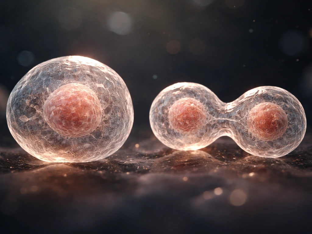
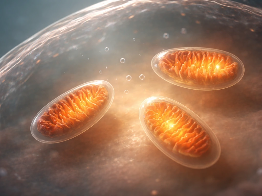
But growth also serves a forward-looking purpose. Before a cell can divide, it needs enough raw material to produce two complete, functional daughter cells. That means doubling its ribosomes, duplicating its mitochondria, replicating its entire genome, and building enough membrane to wrap two separate cells. If a cell tried to divide without first accumulating all that biomass, it would produce two undersized, under-equipped daughters that would probably die. So growth is the necessary prep work for reproduction. Do cells grow continuously or only in bursts? In most cell types, growth happens in distinct phases tied to where the cell sits in its division cycle, not as a constant, uninterrupted process.
There's also a metabolic throughput argument. As a cell takes on more work, responding to hormones, secreting proteins, repairing DNA, it needs more molecular machinery. A liver cell processing a flood of glucose after a meal literally needs more enzymes to handle the load. Growing in size is the cellular equivalent of hiring more staff and buying more equipment.
What tells a cell to grow: signals, nutrients, and self-monitoring
Cells don't just grow whenever they feel like it. They listen carefully to their environment and to internal status signals before committing resources to growth. The main inputs are nutrient availability, energy status, oxygen levels, and external growth factors, chemical signals from other cells that say, essentially, "we need more of you right now."
The most important molecular hub connecting all these signals is the mTOR signaling network (mechanistic target of rapamycin). When nutrients and growth factors are plentiful, mTOR complex 1 (mTORC1) switches on a broad anabolic program: it ramps up protein synthesis, promotes lipid synthesis for new membranes, and suppresses autophagy (the cell's internal recycling system). In practical terms, mTOR is the master "grow now" switch. When the environment is favorable, mTOR pushes resources toward building biomass.
Flip the situation around and you see the brake system just as clearly. When energy is low or oxygen is scarce, a sensor called AMPK activates. AMPK then activates the TSC complex, which inhibits mTOR signaling directly. The result: growth programs get paused, anabolic activity slows, and the cell shifts toward conservation and recycling instead of building. This AMPK-TSC-mTOR axis is essentially a cellular energy meter, it prevents a cell from spending resources it doesn't have.
Growth factors add a social layer to this decision. Signals like EGF (epidermal growth factor), insulin-like growth factors, and many others bind to surface receptors and activate intracellular cascades (including the PI3K/Akt pathway, which feeds into mTOR). These signals tell a cell that the organism as a whole needs more cells in a particular tissue. Without them, most cells default to a resting, non-growing state rather than growing autonomously.
Growing bigger vs. dividing: how the cell cycle connects both
Cell growth and cell division are related but distinct events, and understanding which is happening at any moment requires knowing where a cell is in its cycle. How cells grow or increase in size is actually a separate question from how they divide, both matter, but the biology behind each is different.
The cell cycle has four main phases: G1, S, G2, and M. During G1 (first gap phase), the cell grows in size, it synthesizes proteins, expands its organelles, and assesses whether conditions are right to commit to division. This is where most of the "growth" in the casual sense happens. The S phase is dedicated to DNA replication. G2 is a second growth and preparation phase where the cell double-checks its replicated DNA and assembles the machinery for division. M phase (mitosis) is the actual split, the cell divides its duplicated chromosomes into two nuclei and then physically pinches into two daughter cells.
Checkpoints guard the transitions between phases. The G1 checkpoint (sometimes called the restriction point in mammalian cells) is the most critical: if the cell passes it, it's committed to dividing. The G2/M checkpoint checks that DNA replication is complete and error-free. The G2-M (DNA damage) checkpoint behavior includes p53-dependent arrest and is mechanistically tied to blocking transitions into S or mitosis via checkpoint pathways [p53 can halt the cycle](https://aacrjournals.
org/clincancerres/article/8/11/3311/289049/To-Arrest-or-Not-To-G2-M-Cell-Cycle). If damage is detected, checkpoint proteins like p53 can halt the cycle to allow repairs, or trigger cell death if the damage is too severe. These checkpoints are why normal cells don't divide recklessly. They are, in essence, quality-control inspections.
How a cell grows through each of these phases depends heavily on whether it's passed each checkpoint successfully.
Why cells can't keep growing forever: the hard physical and biochemical limits
Here's a question worth sitting with: if growth is so important, why don't cells just keep getting bigger instead of dividing? The answer is physics, and it's one of the more elegant constraints in all of biology.
The surface area to volume problem
As a cell gets larger, its volume grows much faster than its surface area. Volume scales with the cube of the radius; surface area scales with the square. This matters enormously because the cell membrane is the only interface through which nutrients can enter and waste products can exit. A cell that doubles in radius has 4 times as much surface area but 8 times as much volume to supply.
At some point, the membrane simply can't move nutrients in and waste out fast enough to keep the interior alive. [Diffusion, the passive movement of molecules across distances](https://bio. libretexts. org/Bookshelves/IntroductoryandGeneralBiology/Book%3AGeneralBiology%28Boundless%29/33%3ATheAnimalBody-BasicFormand_Function/33.
04%3AAnimalFormandFunction-LimitingEffectsofDiffusiononSizeandDevelopment), also becomes the limiting factor: small molecules like oxygen can only diffuse effectively across distances of roughly 10 to 20 micrometers before concentration gradients become too shallow to drive meaningful transport. Most animal cells stay in the 10 to 30 micrometer range for exactly this reason. Do cells get bigger as you grow as an organism? In many cases, it's actually cell number that increases rather than individual cell size.
Biochemical and molecular crowding limits
Even if you could solve the diffusion problem, there are biochemical ceilings. A single copy of the genome can only be transcribed so fast. A finite number of ribosomes can only produce so many proteins per minute. As a cell grows, the ratio of DNA to cytoplasm can become limiting, the genome simply can't produce mRNA fast enough to direct protein synthesis for a much larger cell. Additionally, the interior of a cell is already extremely crowded. Proteins, RNA, organelles, and metabolites pack the cytoplasm so densely that transport and signaling become sluggish when cell volume expands beyond manageable limits.
Energy and waste constraints
Larger cells need more ATP. Mitochondria (or, in bacteria, the membrane itself) are the power plants, and they have to scale with cell volume. Producing ATP requires oxygen, and getting oxygen to the cell's core becomes harder as size increases. On the waste side, carbon dioxide and metabolic byproducts need to escape. A very large cell risks accumulating toxic waste faster than it can remove it. Division solves all of these problems neatly: two smaller cells each have a more favorable surface area to volume ratio, better diffusion distances, and a genome-to-cytoplasm ratio that makes sense.
Growth across tissue types: it's not one-size-fits-all
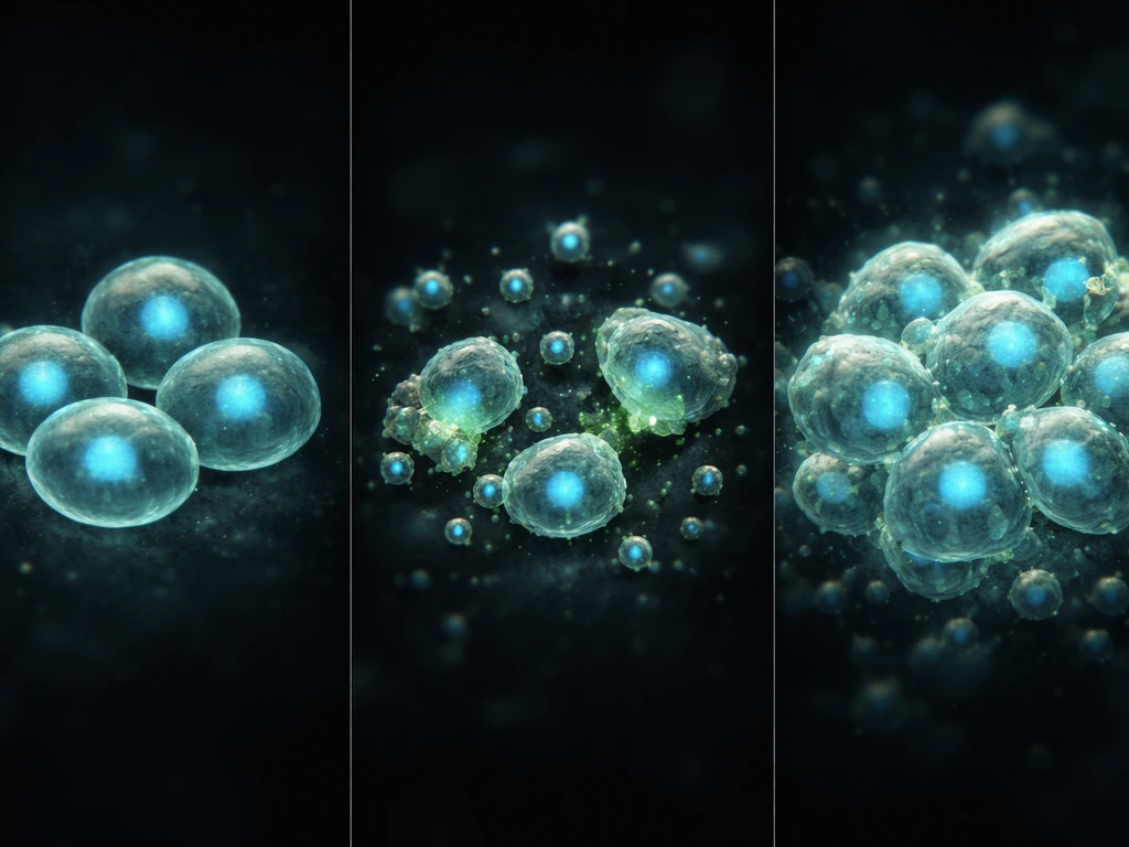
Different tissues use growth in remarkably different ways. Understanding what two processes tissues use to grow gets at a fundamental distinction: tissues can expand either by having individual cells get larger (hypertrophy) or by increasing cell number through division (hyperplasia). Most tissues use a combination of both, but the balance varies a lot depending on the tissue type and the organism's developmental stage.
Epithelial tissues, for example, grow primarily through rapid cell division rather than enlargement. How epithelial tissue grows is tightly regulated by signals from the basement membrane and neighboring cells, with contact inhibition playing a major role in stopping growth when the tissue reaches the right size. Adipose tissue works quite differently: fat cells (adipocytes) can expand dramatically in volume as they accumulate lipid droplets, representing a clear case of hypertrophic growth. Adipose tissue can grow through both cell enlargement and the creation of new adipocytes from precursor cells adipose tissue grows. <a data-article-id="D9AE1F87-5E5A41E3-AFCB-598843C138AF">How adipose tissue grows also involves recruiting precursor cells and converting them into new adipocytes, so it mixes both hypertrophy and hyperplasia depending on conditions.
The skin is a particularly interesting example when it comes to thinking about which cells are doing the dividing. Which cells in the epidermis grow and divide are the stem-like basal cells sitting at the deepest layer, not the flattened, keratinized cells at the surface. The basal cells divide continuously, pushing daughter cells upward where they differentiate and eventually shed. This is a beautiful example of how growth is spatially and functionally organized in a tissue: only specific cells in specific locations carry out active division.
Simple ways to observe and test cell growth yourself
You don't need a cutting-edge research lab to build real intuition for cell growth. Even at a teaching lab or community college level, a few conceptual experiments reveal the core principles very clearly.
- Growth curves with yeast or bacteria: Inoculate a culture in rich media, take optical density readings every 30 to 60 minutes, and plot the curve. You'll see a lag phase (cells adapting), an exponential phase (rapid growth and division), a plateau (resource depletion or waste accumulation), and decline. Change one variable — reduce glucose, remove a nitrogen source, drop temperature — and watch how each phase shifts. This directly illustrates how nutrient availability drives growth rate.
- Serum starvation and growth factor re-addition: Mammalian cell cultures can be driven into a non-dividing resting state by removing serum (which contains growth factors). Adding serum back triggers re-entry into the cell cycle. Even observing colony density before and after under a simple light microscope makes the growth-factor dependence tangible.
- Oxygen and crowding effects: Compare a culture grown in a standard incubator with one that's significantly overpopulated (too many cells per dish). The crowded culture will slow and stop dividing due to contact inhibition and oxygen/nutrient depletion — a physical demonstration of the constraints described above.
- Microscopy of dividing cells: Under a phase-contrast or basic light microscope, actively dividing cells in culture will show rounded-up morphology during mitosis, visible chromosome condensation (if stained), and physical pinching (cytokinesis). Counting the proportion of cells in division (mitotic index) gives a direct read on how actively a population is growing.
- Agar diffusion analogy: Press a small paper disc soaked in a nutrient into a thin agar gel and watch diffusion gradients stain over time. This is a useful analog for understanding why diffusion limits cell size — nutrients can only penetrate so far before concentration drops below usable levels.
The key habit to build is connecting environmental variables (nutrients, oxygen, growth factors, crowding) to measurable outputs (growth rate, division frequency, cell size). Once you can reason about that chain intuitively, most cell biology questions about growth start answering themselves.
When growth stops or goes wrong
Normal cells don't grow indefinitely. They have sophisticated mechanisms for parking, reversing course, or eliminating themselves when conditions demand it.
Quiescence: the deliberate pause
Many differentiated cells in the body exist in a state called G0, they've exited the active cell cycle and are not dividing. This isn't death or damage; it's a programmed resting state called quiescence. Neurons, muscle cells, and many stem cells spend most of their lives in G0. They can re-enter the cell cycle if the right signals arrive (wound healing, tissue damage, growth factor stimulation), but their default is to stay put. Quiescence is actually a sophisticated, actively maintained state, cells in G0 have altered gene expression patterns compared to cycling cells and have reduced metabolic activity.
Apoptosis: programmed self-destruction
When a cell detects irreparable DNA damage, viral infection, or loss of survival signals, it can trigger apoptosis, a clean, controlled self-destruction. Apoptosis is not the same as necrosis (messy cell death from injury). In apoptosis, the cell packages its contents into membrane-bound fragments that neighboring cells and immune cells can safely engulf and recycle. This sounds grim, but it's critical for development (it's how the spaces between your fingers formed in the embryo) and for preventing damaged cells from becoming dangerous. The p53 protein is a central decision-maker here: it senses DNA damage and can trigger either cell-cycle arrest (to allow repair) or apoptosis (if repair isn't possible).
Runaway growth: when the brakes fail
Cancer is fundamentally a failure of the growth control systems described in this article. Mutations in oncogenes (which promote growth) or tumor suppressors (which restrain it) allow cells to grow and divide without proper signals, ignore checkpoint warnings, avoid apoptosis, and sometimes override contact inhibition. The result is uncontrolled proliferation. Interestingly, many cancer cells show hyperactive mTOR signaling, the growth accelerator is stuck in the "on" position even without external growth factors. This is why mTOR has become a target for cancer therapies. Understanding why normal cells stop growing is, in many ways, the most direct path to understanding why cancer cells don't.
Stress responses and adaptive growth changes
Cells under nutrient stress, hypoxia, heat shock, or oxidative damage don't just divide or die, they activate a spectrum of stress-response programs. The AMPK-mTOR axis mentioned earlier is one. The unfolded protein response (UPR) handles the accumulation of misfolded proteins in the endoplasmic reticulum. Autophagy (that cellular recycling system mTORC1 suppresses during good times) ramps up under stress to break down damaged organelles and recycle amino acids. These stress responses can temporarily shrink cell size, reduce proliferation rate, and redirect energy toward survival rather than growth. They're adaptive and reversible, when conditions improve, growth programs resume.
The full picture: a quick comparison of growth states
| Growth State | Key Signal | mTOR Status | Outcome |
|---|---|---|---|
| Active growth (G1/S/G2) | Nutrients + growth factors abundant | Active (mTORC1 on) | Protein/lipid synthesis, cell enlargement, DNA replication |
| Division (M phase) | G2/M checkpoint passed | Moderately active | Chromosome segregation, cytokinesis, two daughter cells |
| Quiescence (G0) | Growth factor withdrawal, contact inhibition | Suppressed | Reduced metabolism, no division, reversible pause |
| Stress response | Low energy, hypoxia (AMPK activation) | Inhibited via TSC complex | Growth arrest, autophagy, resource conservation |
| Apoptosis | DNA damage, loss of survival signals | Irrelevant (cell commits to death) | Controlled cell death, safe recycling by neighbors |
| Uncontrolled proliferation (cancer-like) | Oncogenic mutations, constitutive signaling | Constitutively active | Growth without external signals, checkpoint evasion |
Putting it all together
The causal chain for cell growth looks like this: environmental and internal signals (nutrients, growth factors, energy status) feed into molecular hubs like mTOR and AMPK, which dial anabolic programs up or down. When growth is green-lit, cells accumulate biomass through protein and lipid synthesis, expand their organelles, and replicate their DNA. Physical and biochemical limits (surface area to volume, diffusion, genome throughput, energy supply) set the ceiling on how large a cell can get before it's more efficient to divide. Division checkpoints ensure the process is accurate. And when signals are absent, growth stalls into quiescence; when damage is irreparable, apoptosis takes over; when controls fail, uncontrolled proliferation follows.
If you're studying this for a class, focus on the signal-to-outcome chain: what the cell senses, what molecular switch it flips, and what biological consequence follows. If you're designing an experiment, start with one variable (nutrient concentration, oxygen level, serum availability) and measure one output (growth rate, cell count, mitotic index). That's how the underlying logic of cell growth becomes genuinely intuitive rather than just memorized.
FAQ
Do cells always grow before dividing, or can they divide even if growth slows?
Some cells pause growth for long periods without fully leaving the cell cycle, they enter a reversible “brake” state such as G0 or a transient quiescent-like arrest. Others can remain actively growing but slow the rate, for example by downshifting translation and anabolic pathways. The practical clue is whether the cell is still synthesizing DNA or only ramping metabolism and protein turnover.
If nutrients are limited, do cells divide anyway, or will they get stuck later?
In many cell types, size checkpoints are not the same thing as “time in the cycle.” Cells can be driven to G1/S by growth-factor and nutrient cues, even if their size distribution is shifted. However, smaller-than-normal cells often struggle later because they must still complete DNA replication and build division-ready machinery, so downstream checkpoints and stress responses catch up.
What is the difference between a cell getting bigger and a cell actually preparing to divide?
Growth and division are coupled, but they are not identical. A cell can increase in size through G1 growth and organelle expansion without dividing immediately, and it can also increase proliferation markers if replication machinery is activated. The article’s “growth programs green-lit” idea corresponds to anabolic buildup, while checkpoints specifically govern whether the cell will proceed to DNA replication and mitosis.
Why doesn’t high nutrient availability always cause maximum cell growth?
mTORC1 is a strong driver of anabolic growth, but it is not the only control. Cells also integrate signals through pathways such as PI3K/Akt, and they use stress-response routes like AMPK activation and UPR to override growth even when nutrients are present. In other words, “nutrient availability” sets the stage, stress signaling can still veto growth.
Can cells restart growth after hypoxia or oxidative stress, or is growth always permanently halted?
Yes, in some contexts. Cells can recover after stress by resuming translation and membrane synthesis once AMPK activity drops and anabolic signaling returns. But recovery depends on whether damage was repairable, for example severe ER stress that triggers lasting UPR, or irreparable DNA damage that activates p53-dependent arrest or apoptosis.
Is G0 just cells being inactive, or is it a distinct biological program?
Quiescence (G0) is a maintained, gene-expression rewiring state, not a passive “off” switch. Cells typically keep survival programs active, reduce proliferation signals, and often change chromatin organization and metabolic routing so that re-entry into cycling is possible but controlled.
How do cells know when they are crowded, and why does that fail in cancer?
Contact inhibition is tissue-specific, and it depends on how cells sense crowding through adhesion and mechanotransduction, not just membrane area. Some epithelial cancers bypass or weaken contact inhibition, allowing continued growth even when cell density is high.
Which limit becomes dominant first as a cell grows, diffusion, DNA transcription, or energy supply?
As cells get larger, the problem is not only diffusion but also scaling of biogenesis rates, energy production capacity, and waste clearance. Even if nutrients can reach the center, the genome-to-cytoplasm balance and ATP generation must scale with size, otherwise growth stalls or stress pathways ramp up.
If you artificially stimulate growth signaling, will cells grow faster in a straightforward way?
Overexpressing “growth” pathways in culture can sometimes increase size or accelerate cell cycle entry, but it often triggers compensatory stress responses. For example, too-rapid growth without adequate protein-folding capacity can activate UPR, and excess anabolic drive can increase reactive stress that activates apoptosis or senescence.
In experiments, what should I measure to distinguish slowed growth from increased cell death?
A typical practical readout is cell number over time (population expansion) plus a size distribution measure (single-cell imaging or flow cytometry forward scatter). You also need to track viability, because nutrient stress can reduce growth rate while increasing death, which can masquerade as “no growth” if you look only at confluency.
Next Article
What Are Two Processes by Which Tissues Grow
Tissues grow by mitosis to make more cells and by cell enlargement to increase size, guided by signals and limits.