Cells can't grow too large because the physics of diffusion, the geometry of surface area versus volume, and a set of tightly regulated molecular checkpoints all conspire to make a big cell a bad cell. The bigger a cell gets, the harder it is to move nutrients in, push waste out, coordinate chemistry across its interior, and replicate DNA accurately. At a certain point, the cell simply works better if it divides rather than keeps expanding. In general, cell cultures that can grow indefinitely are called immortalized cell lines. That's not a rule anyone wrote down, it's a constraint baked into the laws of physics and refined over billions of years of evolution.
Why Can’t T Cells Grow Too Large? Size Limits Explained

Marcus Whitmore
19 Apr 2026
Cell growth basics: size vs. dividing at the right moment
Growth and division are not the same thing, and that distinction matters a lot here. A cell grows by synthesizing new proteins, lipids, and organelles, which increases its mass. At some point, it divides, which splits that mass between two daughter cells. The question is: why doesn't the cell just skip the division step and keep piling on mass indefinitely? This is exactly the size-and-division problem the cell cycle solves by enforcing division at the right point why cells don't just continue to grow larger.
The short answer is that cells have evolved size-sensing mechanisms that actively couple growth to division. In budding yeast, for example, there's a commitment point in the cell cycle called Start, sitting at the G1/S boundary. The cell will not progress to DNA replication until it has reached an appropriate size threshold. It's not drifting into division, it's waiting until a size signal is strong enough to flip the switch.
In human cells, something similar happens through inhibitor dilution. The cell-cycle inhibitor Rb is present at a fixed amount at the start of G1. As the cell grows and its volume increases, the concentration of Rb drops because the same amount of protein is spread across a larger cell. When Rb concentration falls below a critical level, the brakes come off and the cell commits to division. This means growth itself is the trigger for division, which elegantly prevents a cell from growing indefinitely without check.
Diffusion and surface area-to-volume: the geometry problem
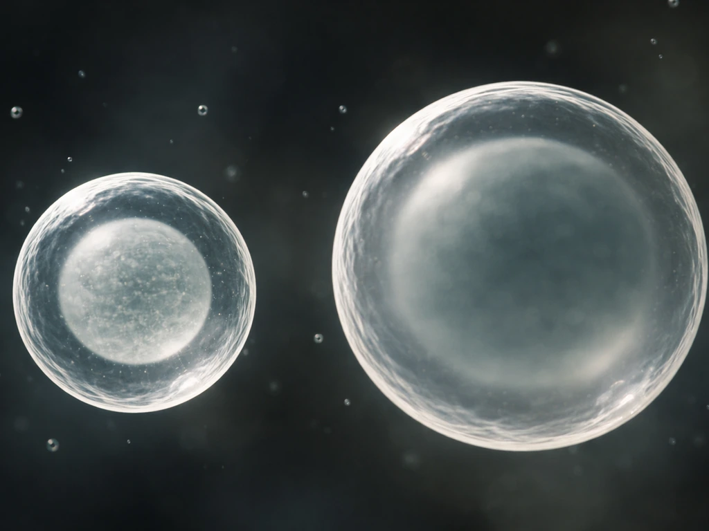
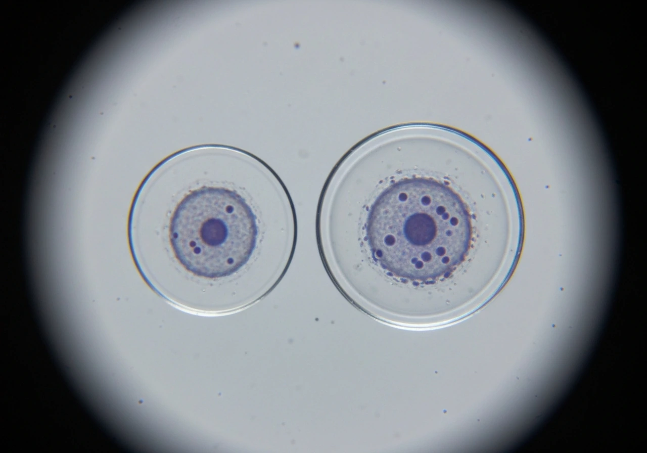
This is the most fundamental physical reason cells can't grow without limit, and it's worth spending a moment with the math to build real intuition. Imagine a cube. When you double its side length, the surface area goes up by a factor of 4, but the volume goes up by a factor of 8. The ratio of surface area to volume drops by half. Now think about what that means for a cell: the surface (the membrane) is where everything enters and exits. The volume is everything that needs to be fed and cleaned up.
Diffusion is the only way many molecules move across short distances inside a cell, and it's exquisitely fast at small scales but painfully slow at large ones. Diffusion time scales with the square of the distance. Double the cell's radius, and molecules take four times as long to reach the center. A small bacterial cell might be 1 to 2 micrometers across, a size where diffusion works just fine. A large eukaryotic cell might be 10 to 100 micrometers across. At 100 micrometers, passive diffusion to the center is tens of thousands of times slower than at 1 micrometer.
The practical result is that as a cell grows larger, its surface area becomes proportionally smaller relative to its interior demands. There's simply not enough membrane to ferry in the nutrients and signal molecules the growing interior needs, and not enough exit points to handle the waste being generated. The cell hits a transport ceiling.
Nutrients, oxygen, and waste removal: the supply chain breaks down
Think of the cell's interior like a city. A small town can thrive with a few roads in and out. A megacity with the same number of roads would be gridlocked. As cell volume increases, the metabolic demand for oxygen and glucose scales with the interior mass, but the ability to supply those resources scales only with the surface area. This mismatch gets worse and worse as the cell grows. In other words, populations also fail to grow without limit when their resource supply cannot keep up with increasing demand resource supply scales with surface area while demand scales with volume.
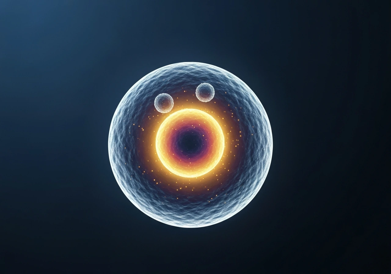
Oxygen is the tightest constraint for aerobic cells. It diffuses passively across the membrane and through the cytoplasm, and it gets consumed rapidly by mitochondria. In a very large cell, oxygen levels at the center would drop dramatically because it is consumed faster than it can diffuse inward. This creates a hypoxic core, which is actually what happens in large tumors, where the center dies due to oxygen starvation. A healthy cell avoids this fate by staying small enough that diffusion keeps the interior supplied.
Waste removal is the flip side of the same problem. Metabolic byproducts like carbon dioxide, ammonia, and excess ions must be exported continuously. In a small cell, these molecules reach the membrane quickly and exit. In a large cell, they accumulate in the interior faster than they can diffuse out, disrupting the local pH, ionic balance, and enzymatic conditions that cellular chemistry depends on. The cell's own metabolism would poison it.
Internal organization limits: crowding, the cytoskeleton, and error rates
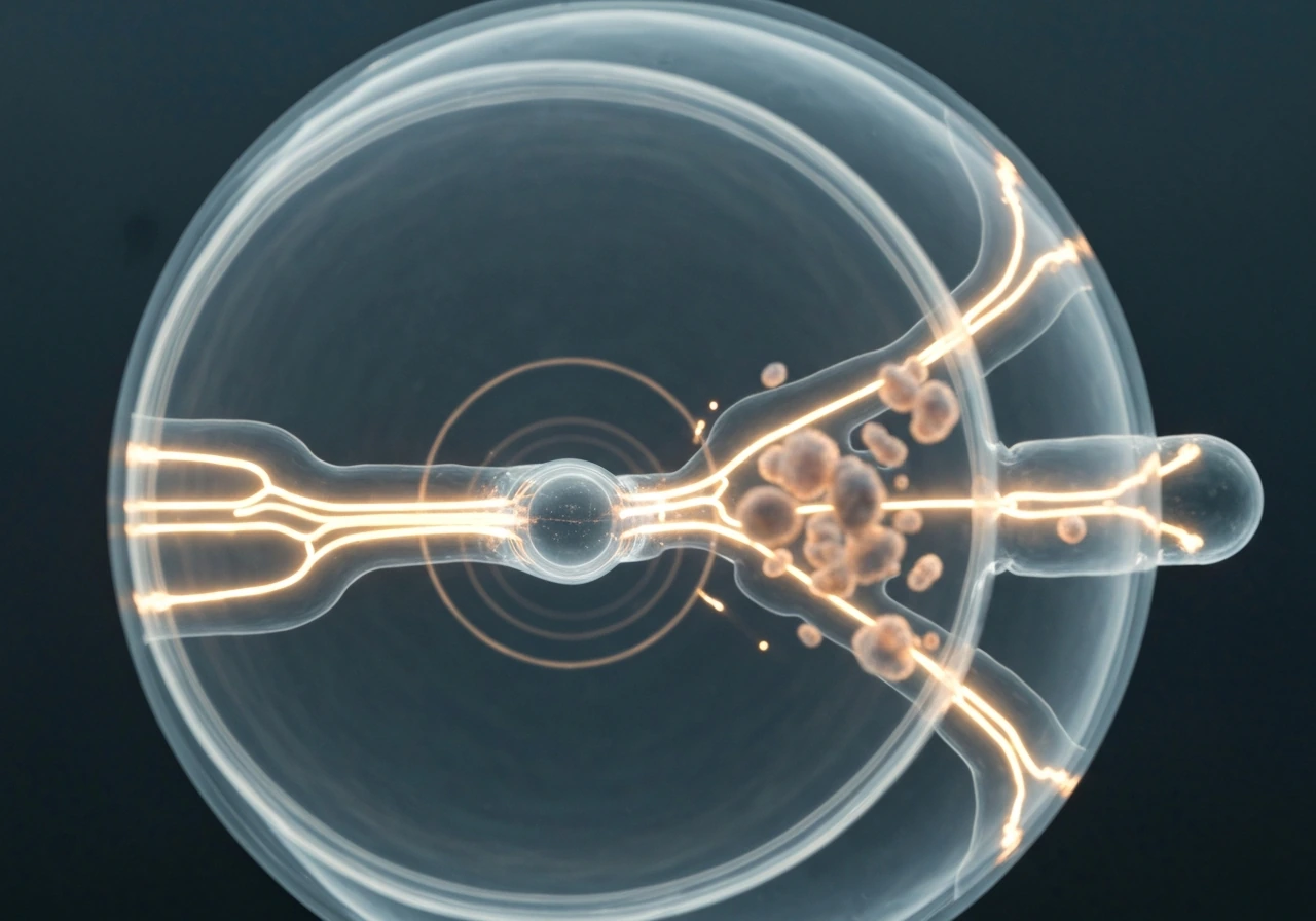
Even if you could magically solve the diffusion problem, a very large cell would still run into serious internal organization issues. The cell's cytoskeleton, a network of protein filaments including actin, microtubules, and intermediate filaments, provides the structural scaffolding that organizes the interior, positions organelles, and drives intracellular transport. As cell volume grows, maintaining and coordinating that scaffolding across larger distances becomes increasingly difficult and energetically expensive.
Macromolecular crowding is another underappreciated constraint. The cytoplasm is already extraordinarily dense, packed with proteins, RNA, ribosomes, and metabolites at concentrations that would seem impossible by laboratory standards. As a cell gets larger, keeping that molecular density consistent across the whole volume requires proportionally more resources. Regions of the cell become poorly mixed and chemically inconsistent, meaning the same enzymatic reaction can proceed at different rates in different parts of the cell. That's a reliability disaster for a system that depends on tight biochemical regulation.
DNA replication accuracy is also at stake. The larger the cell, the more mass must be coordinated and the longer replication takes, increasing the window for errors. The genome can only be copied at a fixed rate, so a very large cell would take much longer to replicate its DNA, increasing replication errors and the chance of mutations. Cell-cycle checkpoints exist partly to prevent this from spiraling out of control.
Why cell-cycle checkpoints actively block runaway growth
The cell doesn't just passively run into physical limits. It has active molecular machinery that detects when size thresholds have been reached and enforces division. This is one of the most elegant aspects of cell biology, the cell polices its own size.
In mammalian cells, the mTOR pathway (along with its partners PI3K, S6K1, and 4EBP1/eIF4E) is a central coordinator of cell growth and cell-cycle progression. This is consistent with reviews of mTOR signaling showing that mTOR integrates nutrient, growth factor, and energy cues to regulate cell growth and coordinate entry through the cell cycle mTOR pathway is a central coordinator of cell growth and cell-cycle progression. mTOR senses nutrient availability, growth factor signals, and energy status, then calibrates the rate of protein synthesis and ribosome production accordingly. It also communicates with the cell-cycle machinery to coordinate when the cell has grown enough to divide. If mTOR signaling is artificially overactivated, cells grow excessively, which is relevant to understanding cancer biology.
There is also a p38 MAPK-dependent size checkpoint in animal cells that regulates the length of G1. This checkpoint actively allows cells that have grown large enough to proceed into S phase, while holding smaller cells back. Think of it as a height requirement for getting on a ride, except the ride is DNA replication. Research has shown that this checkpoint maintains size uniformity across a cell population, actively correcting for cells that start G1 at an unusually large size by shortening their G1 phase.
The takeaway is that unlimited growth isn't just physically impractical, it's also actively suppressed by the cell's own regulatory networks. When those networks malfunction and size control breaks down, the consequences are severe, which is closely related to why uncontrolled cell growth is the defining feature of cancer.
What sets the 'too large' threshold for different cell types
The exact size limit isn't the same for every cell. It shifts based on the cell's metabolic rate, the available transport machinery, and the specific tasks the cell performs. High-metabolism cells like liver cells (hepatocytes) or muscle cells that are burning energy constantly have a tighter effective size limit because they consume oxygen and generate waste faster. Slower-metabolism cells can tolerate slightly larger sizes before diffusion becomes the limiting factor.
Cells that have specialized transport infrastructure can push the envelope. Neurons are a great example: a single neuron can have an axon that extends meters in length, which seems to violate everything discussed above. The trick is that axons are extremely thin tubes, so they maintain a favorable surface area-to-volume ratio along their length. The cell body itself stays compact. Active transport systems like motor proteins (kinesin and dynein) shuttle cargo along microtubules rather than relying on diffusion alone.
Multinucleate cells represent another workaround. Some large cells, like skeletal muscle fibers or the syncytiotrophoblasts of the placenta, contain multiple nuclei distributed throughout their volume. Each nucleus effectively governs a local territory, reducing the distance between genetic control and the regions that need it. This is a biological acknowledgment that the single-nucleus model breaks down at large sizes.
| Cell type | Approximate size | How it handles size limits |
|---|---|---|
| E. coli (bacterium) | 1–2 µm | Stays tiny; relies purely on diffusion; divides rapidly |
| Typical human cell | 10–20 µm | Active transport, organized cytoskeleton, size checkpoints at G1/S |
| Neuron cell body | ~20 µm body, meter-long axon | Thin axon keeps SA:V ratio high; motor proteins replace diffusion along axon |
| Skeletal muscle fiber | Up to cm in length | Multinucleate; multiple nuclei share governance of large volume |
| Ostrich egg yolk | ~10 cm | Metabolically nearly inactive; yolk is stored nutrient, not living cytoplasm |
| Purkinje neuron | ~70–80 µm body | Highly branched dendrites; each branch stays thin for diffusion efficiency |
Real-world examples and what they teach us
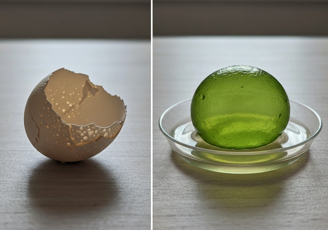
The ostrich egg is the largest single cell you can hold in your hands, and it's the perfect illustration of why 'large cell' is a misleading concept. The massive yolk is not metabolically active cytoplasm in the way a liver cell is. It's essentially stored nutrient material, inert fat and protein packed in by the mother. The actual living portion of the egg is a tiny disc of cells sitting on top of the yolk. Nature achieved 'large' by cheating: it filled the volume with non-living material rather than scaling up living chemistry.
Valonia ventricosa, a single-celled green alga sometimes called the 'sailor's eyeball,' can reach 5 centimeters in diameter and is often cited as the world's largest single-celled organism. It survives at this scale by maintaining a large central vacuole (essentially a bag of water and ions) that occupies most of the cell's volume, with the actual living cytoplasm pushed into a thin layer near the cell membrane. Again, nature achieves large size by keeping the metabolically active living volume thin and close to the membrane.
Most multicellular organisms bypass cell-size limits entirely by using many small cells rather than a few large ones. A human body contains roughly 37 trillion cells, each staying small enough for diffusion and transport to function efficiently. This strategy, many small units working together, is almost certainly why multicellularity evolved and was so successful. It's not an accident that complex life is built from cells in the 10 to 100 micrometer range.
How to reason about cell-size limits practically
If you're a student trying to reason through or model cell-size constraints, here are the key variables to think about:
- Surface area-to-volume ratio: calculate this first for any cell shape. Sphere volume = (4/3)πr³; surface area = 4πr². As r increases, SA:V decreases as 3/r. This ratio alone tells you a lot about diffusion capacity.
- Diffusion distance and rate: diffusion time scales with distance squared (t ≈ x²/2D). Doubling the cell radius quadruples the time for molecules to reach the center.
- Metabolic rate: a high-metabolism cell hits the wall sooner than a low-metabolism cell at the same size. Compare oxygen consumption rates when modeling.
- Active transport infrastructure: does the cell have motor proteins, a well-organized cytoskeleton, or specialized channels? These can extend the effective functional size.
- Nuclear-to-cytoplasmic ratio: as cell volume grows without more nuclei, the nucleus must govern more cytoplasm, which eventually limits the cell's ability to coordinate gene expression.
- Cell-cycle checkpoint signals: mTOR signaling, Rb dilution, and p38 MAPK activity are the key molecular handles for understanding when the cell 'decides' it is large enough to divide.
Conditions that shift the threshold include temperature (higher temperatures speed up diffusion and metabolism but also increase error rates), oxygen availability (lower oxygen tightens the limit for aerobic cells), and cell shape (elongated or flattened cells maintain better SA:V ratios than spheres of the same volume, which is why red blood cells are biconcave discs rather than spheres).
If you're exploring related questions, you might also be curious about what sets limits on indefinite cell growth more broadly, or what conditions allow certain cell lines to bypass normal size and division controls entirely. In this context, the main reason cells cannot grow indefinitely comes down to size-dependent physical limits and active cell-cycle checkpoints that enforce division why can't cells grow indefinitely. Those questions connect directly to topics like cell-cycle regulation, contact inhibition, and why most normal cells in your body divide a finite number of times before stopping. The size question you started with sits at the center of all of it.
FAQ
Can a T cell divide without first getting bigger, or does it always need to grow past a size threshold?
It generally needs to pass size-related checkpoints, but the trigger is not just absolute size. Nutrient and signaling strength (for example through mTOR and growth-factor pathways) can accelerate G1 progression once size and conditions are sufficient. In practice, cells that start G1 too small often take longer to reach the commit point or fail to enter S phase efficiently.
Do T cells have a single fixed “maximum size,” or is the limit different depending on the situation?
There is no universal maximum. The effective size limit shifts with oxygen availability, metabolic rate, nutrient access, and the cell’s geometry. A T cell encountering richer growth conditions can sustain a larger metabolically active volume before diffusion and waste-removal constraints dominate.
Why do activated T cells sometimes appear to get larger before dividing?
Activation increases biosynthesis and metabolic demand, so the cell expands its mass and replication machinery. The growth is then coupled to division via G1/S control, so once growth indicators (like inhibitor dilution and checkpoint signaling) cross a threshold, the cell commits to DNA replication rather than continuing to expand.
If diffusion and waste removal are the main reasons, why can some cells be very large in the body?
Many “large” cells are large because their function reduces the burden on intracellular transport. Examples include multinucleate cells (multiple nuclei reduce control distances), specialized shapes that maintain favorable surface area-to-volume ratios, or cases where most volume is non-cytoplasmic storage rather than metabolically active material.
How does cell shape affect the answer to why T cells cannot grow too large?
Shape changes the surface area-to-volume relationship. Elongated or flattened geometries maintain better transport capacity than a spherical cell at the same volume. If a T cell’s shape changes during activation, that can partially mitigate transport limits while it prepares to divide.
What happens to T cells if the size checkpoints are disrupted?
If size control and cell-cycle coupling fail, cells can push into S phase despite being unable to maintain favorable intracellular conditions, increasing replication stress, error rates, and dysfunctional growth. This is one reason checkpoint malfunction is linked to uncontrolled proliferation and cancer-like behavior.
Do temperature changes alter the practical size limit for T cells?
Yes. Higher temperature speeds diffusion and metabolism, which can initially help transport but also increases the rate of biochemical reactions and can raise error risk during replication. So the net effect on the “effective size limit” depends on whether transport improvements outpace increases in stress.
Can T cells grow indefinitely in culture if conditions are optimized?
Normal primary T cells typically do not grow indefinitely even with good media. Immortalization requires additional changes that bypass or disable normal regulatory controls, not just improved nutrients. Optimizing conditions can increase how fast and how many times they divide, but it usually does not remove size-dependent constraints permanently.
Is the oxygen limit relevant to T cells the same way it is for large tumors?
Oxygen matters, but the severity depends on diffusion distances and metabolic demands. T cells are often smaller than the diffusion scales seen in tumor cores, so they rarely face the same degree of hypoxic center. Still, low oxygen environments can constrain proliferation and push cells toward slower growth or altered behavior.
Why is “growth” so closely tied to “division” in T cells, and what does that mean experimentally?
Growth adds mass by building proteins, lipids, and organelles, while division redistributes that mass. Experimentally, you often see that blocking protein synthesis or growth signaling reduces entry into S phase even if the cell has not fully stalled in size, because the cell-cycle machinery expects growth indicators to reach a threshold.
Next Article
What Limits How Large a Cell Can Grow
Explains why cells stop growing: surface-area limits, diffusion of oxygen and wastes, and cell-cycle and structural cons