Animal cells grow in two distinct ways: they get physically bigger by building more proteins, membranes, and organelles during interphase, and they increase in number by completing the cell cycle through DNA replication and division via mitosis and cytokinesis. Both processes are tightly regulated. A cell doesn't just randomly swell and split, it follows a precise sequence of checkpoints, fueled by nutrients and growth signals, and stops when conditions say stop.
How Do Animal Cells Grow: Cell Cycle, Signals, Limits
Marcus Whitmore
14 May 2026
What 'growth' actually means for an animal cell
When biologists talk about animal cell growth, they usually mean one of two things, or both at once. The first is an increase in cell size: the cell synthesizes more protein, builds more membrane lipids, duplicates its organelles, and physically enlarges. The second is an increase in cell number: one cell completes the cell cycle and divides into two daughter cells. In a developing embryo or a healing wound, both things happen simultaneously and in a coordinated way. A liver cell responding to tissue damage, for example, first grows larger to ramp up biosynthetic capacity, then divides to replace lost cells.
This two-part definition matters because it clarifies a common point of confusion. 'Growth' isn't just division, and it isn't just getting bigger. A muscle fiber that adds more contractile protein without dividing is growing. A tumor cell that divides rapidly without much size increase is also growing, but in a very different and dangerous way. Understanding both sides gives you the full picture.
The cell cycle from start to finish

Think of the cell cycle as a project timeline with mandatory review meetings before you're allowed to move forward. The cycle has two broad phases: interphase (the long preparation period) and M phase (the actual division event). Most of a cell's life is spent in interphase, which itself breaks into three sub-stages: G1, S phase, and G2.
- G1 (Gap 1): The cell grows in size, synthesizes proteins, and checks whether conditions are right to commit to division. This is the most variable phase — cells can linger here for hours or years depending on the tissue.
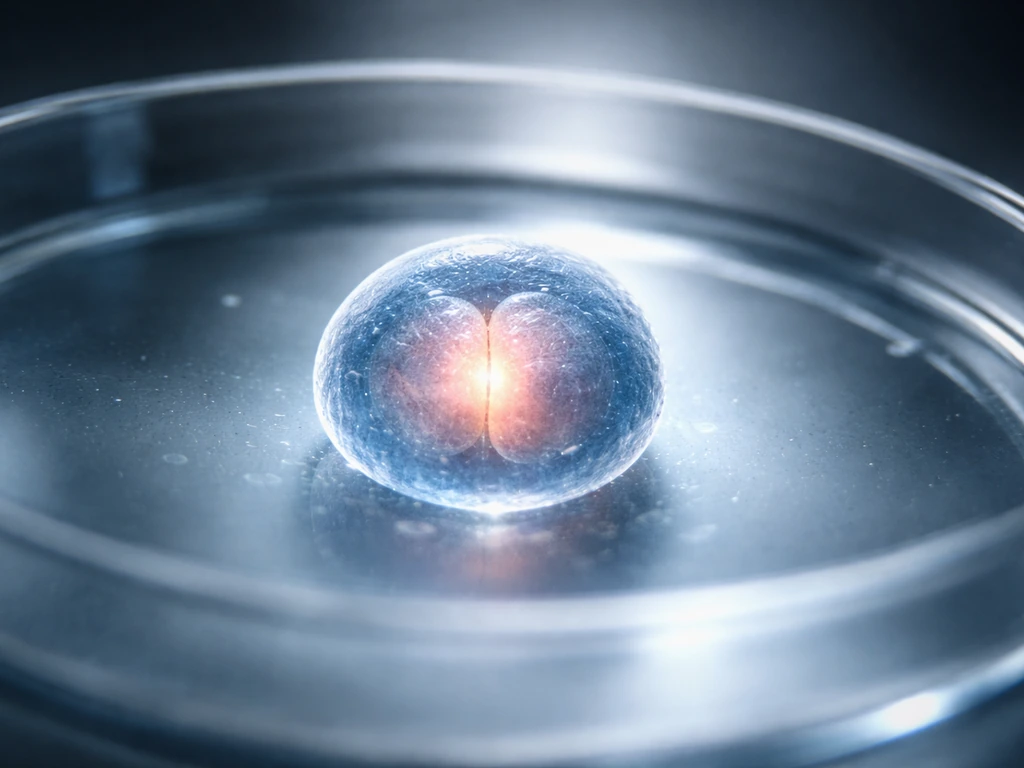
- S phase (Synthesis): DNA replication happens here, along with centrosome duplication. By the end of S phase, the cell has two complete copies of its genome.
- G2 (Gap 2): More protein synthesis and growth, plus final checks that DNA replication completed without errors before the cell enters mitosis.
- M phase: Mitosis separates the duplicated chromosomes into two nuclei, and cytokinesis splits the cytoplasm to produce two daughter cells.
What drives the transitions between stages? Proteins called cyclins team up with enzymes called cyclin-dependent kinases (CDKs). Cyclin levels rise and fall at specific points in the cycle, and when a cyclin binds its CDK partner, it activates the kinase to trigger the next stage. Think of cyclins as the access badges that let CDKs through the door at each checkpoint. Without the right badge, the door stays shut.
The three checkpoints you need to know
There are three major checkpoints in the mammalian cell cycle, and each one is essentially asking the same question: 'Is it safe to continue?' At the G1/S checkpoint, the cell checks for DNA damage and whether growth signals are present. Cyclin D pairs with CDK4/6, and cyclin E pairs with CDK2, to relieve the brake protein Rb from blocking a transcription factor called E2F. Once E2F is free, it activates genes needed for S-phase entry. If DNA is damaged, the tumor suppressor p53 gets activated, which in turn ramps up a CDK inhibitor called p21, that jams the cyclin-CDK machinery and arrests the cell before it can replicate damaged DNA.
The G2/M checkpoint asks whether DNA replication finished correctly before the cell tries to divide. If there are unrepaired breaks or incomplete replication forks, checkpoint signaling blocks entry into mitosis. Then there is the spindle assembly checkpoint (SAC) inside M phase itself: before sister chromatids can separate, every chromosome must be properly attached to spindle fibers from both poles of the cell. A protein complex called the mitotic checkpoint complex (MCC) inhibits the APC/C ubiquitin ligase until every kinetochore is attached. Once all chromosomes are aligned at the metaphase plate and attached correctly, MCC releases its grip, APC/C activates, and it tags proteins called securin and mitotic cyclins for destruction, and that is the molecular trigger for anaphase to begin.
How cells actually get bigger: biosynthesis in action
Size increase isn't passive. The cell has to actively build everything it needs before it can divide. During G1 and G2, ribosomes are running constantly, translating messenger RNA into structural proteins, enzymes, and signaling molecules. The endoplasmic reticulum expands, the Golgi apparatus produces more vesicles, and mitochondria replicate so each daughter cell inherits enough energy-generating capacity. Lipids are synthesized to build new membrane, and nucleotide building blocks are stockpiled for the DNA replication that happens in S phase.
All of this biosynthesis demands raw materials. The cell needs amino acids as nitrogen sources for protein building, glucose and other energy sources to power the reactions, vitamins as enzyme cofactors, fatty acids and cholesterol for membranes, and nucleic acid precursors for DNA and RNA synthesis. It also needs inorganic ions like calcium, magnesium, sodium, and potassium to maintain electrochemical balance and support enzyme function. Remove any one of these and biosynthesis stalls, which is exactly why cells stop growing when nutrients run low.
Dividing to multiply: mitosis and cytokinesis step by step
Once a cell passes the G2/M checkpoint, it enters mitosis. Mitosis isn't a single event, it moves through four sequential stages, each with a specific job.
- Prophase: Chromatin condenses into visible chromosomes. The mitotic spindle begins to form from the two centrosomes, which migrate to opposite poles of the cell.
- Metaphase: Chromosomes align at the cell's equatorial plane, called the metaphase plate. Spindle fibers attach to protein structures on each chromosome called kinetochores. The spindle assembly checkpoint is active here, holding the cell in metaphase until every chromosome is properly attached.
- Anaphase: Once the checkpoint is satisfied and APC/C destroys securin and mitotic cyclins, sister chromatids are pulled apart toward opposite poles. The cell elongates. Cytokinesis begins as a contractile ring of actin and myosin starts to pinch the cell membrane inward.
- Telophase: Chromosomes arrive at the poles and decondense. Nuclear envelopes reassemble around each set of chromosomes. The contractile ring continues to tighten.
- Cytokinesis (completion): The cleavage furrow pinches all the way through, splitting the cytoplasm into two daughter cells, each with its own nucleus and a full complement of organelles.
Each daughter cell starts the cycle again in G1, either committing to another round of division or exiting into a resting state called G0 when signals indicate that division is not needed.
What cells need to keep growing
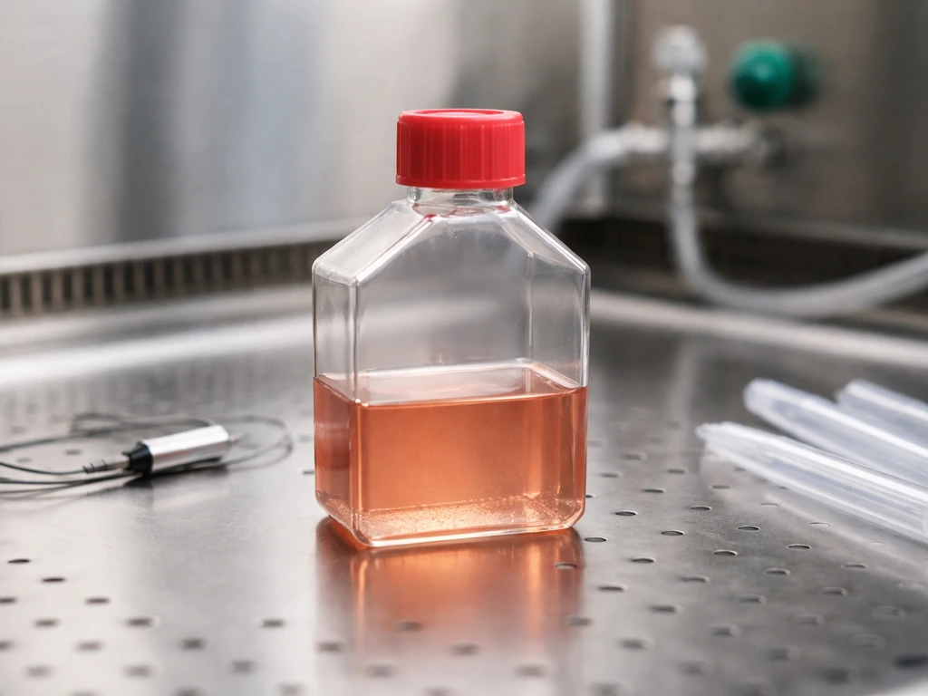
Animal cells are picky about their environment. Get any of the following wrong and growth slows, stalls, or tips into cell death. Lab cell culture makes these requirements very concrete, because researchers have to replicate the right conditions artificially to keep cells alive outside of a body.
| Condition | Optimal range / requirement | Why it matters |
|---|---|---|
| Temperature | 37°C for most mammalian cell lines | Enzyme activity and membrane fluidity are temperature-sensitive; too high or too low disrupts protein function |
| pH | 7.2 to 7.4 | Enzyme catalysis and protein structure depend on tight pH control; deviations stall biosynthesis |
| CO2 / bicarbonate | 5% CO2 with sodium bicarbonate in medium (e.g., 1,500 mg/L in DMEM) | CO2 and bicarbonate buffer the medium to maintain correct pH |
| Oxygen | Adequate supply for aerobic respiration | Mitochondria need O2 to produce ATP; hypoxia forces less efficient anaerobic metabolism |
| Nutrients | Glucose (e.g., 4,500 mg/L in DMEM), amino acids, vitamins, ions, lipids | All biosynthesis reactions require substrate; energy supply (glucose) drives ATP production |
| Cell adhesion substrate | Treated culture surface or extracellular matrix proteins | Many animal cells require surface attachment (anchorage dependence) to receive survival signals and grow normally |
In a real incubator, cells sit in a liquid medium (like DMEM) that supplies glucose, amino acids, inorganic salts, vitamins, and lipid components. Serum is often added as a supplement: it buffers the medium, delivers growth factors and hormones, protects cells from mechanical shear, and helps condition the growth surface of the culture vessel. The incubator holds temperature at 37°C and pumps in 5% CO2 to stabilize pH through the bicarbonate buffer system. It sounds complicated, but it is just recreating the internal environment of a living body.
Why cells don't grow forever
Animal cells have multiple independent mechanisms that put the brakes on growth. This is not a design flaw, it is essential. Unchecked division is cancer. The main constraints are contact inhibition, checkpoint arrest, resource limitation, and apoptosis.
Contact inhibition: crowding sends a stop signal

When healthy epithelial cells touch neighboring cells, surface proteins called E-cadherins form adhesion junctions. These junctions activate the Hippo signaling pathway inside the cell, which phosphorylates a growth-promoting protein called YAP, trapping it outside the nucleus where it cannot stimulate cell-cycle genes. The cell effectively reads the density of its surroundings through its adhesion proteins and downregulates its own proliferation in response to crowding. This contact inhibition is reversible, remove cells from the monolayer (say, by wounding it) and neighboring cells re-enter the cycle to fill the gap. Cancer cells often lose this mechanism, which is why they pile up into tumors.
Checkpoint arrest and DNA damage
If DNA is damaged, by radiation, chemical mutagens, or replication errors, the checkpoint machinery activates p53, which drives expression of p21 and halts cyclin-CDK activity. The cell sits at the G1/S or G2/M checkpoint while repair machinery attempts to fix the damage. If the damage is too severe to repair, the same p53 pathway can redirect the cell toward apoptosis rather than allowing a genetically compromised cell to divide.
Cell size checkpoints
Cells also monitor their own size before committing to division. A cell that hasn't grown large enough in G1 will not efficiently activate the cyclin D/CDK4/6 pathway needed to pass the restriction point and enter S phase. This is one reason nutrient deprivation so reliably arrests cells in G1: without enough building blocks for biosynthesis, the cell never reaches the size threshold that allows cycle progression.
Apoptosis: programmed self-destruction
When stress signals are severe enough, deep DNA damage, loss of survival signals, extreme nutrient deprivation, p53 and other sensors tip the cell into apoptosis, a controlled form of self-destruction. The cell packages itself into small membrane-bound fragments that neighboring cells and immune cells can clean up without triggering inflammation. Apoptosis is not failure; it is the system working correctly to prevent damaged cells from persisting and proliferating.
How scientists study and control animal cell growth
If you want to study how animal cells grow, or if you are designing an investigation, the basic toolkit involves controlling the environment, measuring cell number and health, and probing specific stages of the cycle. Here is how researchers approach it in practice.
Setting up a cell culture experiment
You start by seeding cells at a known density in a flask with complete growth medium. In Excel, you can use text formatting inside cells by enabling word wrap and adjusting row height so longer text displays neatly how to make cells grow with text in excel. Seeding density matters: too sparse and cells may not signal each other properly; too dense and contact inhibition suppresses growth before you can observe it. Temperature stays at 37°C in a humidified CO2 incubator. Variables you can manipulate include nutrient concentration, growth factor levels, serum content, substrate coating, pH, and oxygen tension. Each change lets you isolate which factor is driving or limiting growth.
Measuring viability and proliferation
The simplest readout is a cell count paired with a viability assay. To see whether red blood cells grow, researchers also look at how many immature cells increase and how their viability changes over time. Trypan blue exclusion is the classic method: dead cells take up the blue dye because their membranes are compromised, while live cells exclude it. Count blue (dead) versus clear (alive) cells under a hemocytometer and you get both total number and percent viability at a glance.
To specifically measure cells actively in S phase (replicating DNA), researchers use EdU incorporation. EdU is a modified nucleotide that gets built into newly synthesized DNA. After a pulse of EdU, a click-chemistry reaction tags the incorporated EdU with a fluorescent label, which can then be detected by flow cytometry. This directly tells you what fraction of cells were in S phase during the labeling window, a clean measure of active proliferation rather than just total cell number.
Detecting apoptosis and stress
When you want to know if cells are dying rather than growing, Annexin V staining combined with propidium iodide (PI) is the standard flow cytometry approach. Early apoptotic cells expose a phospholipid called phosphatidylserine on their outer membrane surface, which Annexin V binds. PI enters cells with compromised membranes (late apoptosis or necrosis). Together, the two dyes let you classify cells into live, early apoptotic, late apoptotic, and necrotic populations in a single experiment.
What healthy growth looks like vs. stress
Healthy proliferating cells should show: high viability (typically above 90% in a fresh culture), a predictable doubling time consistent with the cell line's known growth rate, a normal distribution across cell cycle phases by flow cytometry (with a meaningful S-phase fraction), and a monolayer that reaches confluence and then slows, that slowdown is contact inhibition working correctly. Stressed or damaged cells show elevated apoptotic fractions, arrest predominantly in G1 or G2/M on cell-cycle flow plots, and may detach from the culture surface. Tracking these indicators together gives you a comprehensive picture of whether growth is proceeding normally or something in the environment needs adjusting.
For students designing an investigation, the practical next step is to pick one variable (say, glucose concentration or temperature), hold everything else constant, seed cells at the same density across conditions, and measure viability and cell count at 24, 48, and 72 hours. Pair that with an EdU assay if you want to separate 'cells are surviving' from 'cells are actively dividing', those two outcomes do not always go together, and distinguishing them is where the interesting biology lives. Questions about how quickly cells move through the cycle connect directly to factors like how fast do cells grow, while more specialized cases, like why red blood cells don't divide at all in their mature form, reveal just how tissue-specific these growth rules can get.
FAQ
If I see more cells, does that automatically mean the cells were “growing”?
Not always. Cells can increase in size without dividing, for example during early tissue repair when biosynthesis ramps up before mitosis. Conversely, cells can divide rapidly while gaining little net size, which can be a sign of dysregulated growth (for instance, many cancer phenotypes). If you want to know which is happening, measure both cell size (or total protein per cell) and cell division markers like EdU incorporation.
Why can my cell count go up even if proliferation is actually impaired?
Population doubling can hide the details because different fates (division, arrest, death) can offset each other. A fast way to separate outcomes is to pair a proliferation readout (EdU for S phase) with a viability/apoptosis readout (Annexin V/PI or a dye-exclusion assay). This reveals whether the increase is driven by active cycling or by reduced cell loss.
What causes growth to stall if nutrients and temperature seem correct?
Anchorage-dependent cells can stop growing when the substrate is inadequate or when cells detach due to stress. In that case, you may see an apparent drop in proliferation plus a change in morphology, even if nutrients and temperature look correct. To troubleshoot, verify coating type (for example, collagen or fibronectin depending on the cell line), check confluence and detachment, and confirm you are using the correct passaging method and split ratio.
How do I tell if cells are arrested versus just resting (G0)?
Yes, cells can exit the cycle into a resting state (G0) instead of staying arrested in G1 or dying. Nutrient or growth factor withdrawal often pushes cells into a reversible non-proliferative state. Practically, you can distinguish G0 from cycling arrest by looking at S-phase fraction (EdU) and by testing whether reintroducing growth factors and nutrients restores EdU incorporation.
Why does changing serum concentration often make cell growth look inconsistent?
Serum is not only “food,” it also supplies buffering and signaling cues, and it can protect cells from shear stress. Two common pitfalls are using inconsistent serum lots and changing serum concentration between experiments. If your goal is mechanistic, keep serum constant or switch to defined media and then vary only the specific growth factor you want to test.
What are the most common mistakes when measuring S phase by EdU flow cytometry?
Cell cycle measurements require timing and proper gating. With EdU, the labeling pulse length matters, too short a pulse can underestimate S-phase entry, too long can confound interpretation. With flow cytometry, doublets, debris, and aggregates can distort phase distributions. Include doublet discrimination, viability gating, and a properly matched unstained or single-stain control.
How should I choose seeding density so contact inhibition does not ruin the results?
Contact inhibition can make “growth rate” look like it slows down even if the biology is normal. If you seed too high, confluence or crowding will trigger Hippo pathway activity and reduce proliferation before your early timepoints. If you seed too low, you may miss autocrine and paracrine signals. To reduce ambiguity, seed across a narrow density range that stays below full confluence for your entire measurement window.
Can cell cultures have mixed behaviors, like some cells dividing while others die?
Yes. In the same culture, some cells may be arrested in G1 or dying, while a subpopulation continues to cycle. Averaging just total cell count can mask this. Use flow-based fraction measurements (EdU fraction, Annexin V/PI populations) and consider imaging to see whether a resistant subpopulation persists in damaged or low-nutrient conditions.
Does checkpoint arrest always mean the cells will never resume growth?
p53-mediated checkpoints can be transient, cells may pause for repair and then resume cycling if the damage is resolved. If you only measure proliferation at one late timepoint, you might conclude the condition “kills growth” when it actually delays it. A practical approach is to sample multiple timepoints and, when appropriate, use a damage marker or checkpoint-associated readout alongside EdU.
Why don’t mature red blood cells divide like other cells?
Red blood cells are a specialized case where mature cells lack nuclei and do not pass through the DNA replication and mitosis program. However, earlier precursor stages do proliferate. If your experiment involves tissues, remember that “growth rules” differ by developmental stage and cell type, so you need to match the model to the biological question (for example, precursor versus mature erythrocytes).
Next Article
How Fast Do Cells Grow? Typical Rates and What Controls Them
Typical cell growth and division speeds, what controls them, limits on expansion, and how to measure cell cycle rate.