Cell growth and division are controlled by a layered system of molecular signals, internal checkpoints, and physical constraints working together. At the core: growth factors from outside the cell tell it to start preparing to divide, internal cyclin-Cdk protein complexes act like a timed engine driving the cell through each phase, and checkpoint proteins act as brakes that can halt everything if something goes wrong. Nutrients and energy (ATP) set the speed limit, DNA damage can stop the process entirely, and physical size limits cap how fast a cell can realistically grow. When any one of these layers fails, you get uncontrolled proliferation, which is the core logic behind cancer.
What Controls When and How Fast Cells Grow and Divide

Marcus Whitmore
21 May 2026
Growth vs. division, and why timing matters
It's easy to lump cell growth and cell division together, but they're actually separate events that need to be coordinated carefully. But in most cases, cells do not all grow and divide in the same way because they respond differently to growth signals, checkpoints, and physical constraints do all cells grow and divide in the same way.
A cell has to reach a certain size and accumulate enough resources before it can split in two and produce two viable daughter cells. In other words, for most cell types, division generally depends on getting the cell large enough first, so cells do not usually enter mitosis immediately without growth whether cells grow before they divide.
If division happened too fast without adequate growth, each new cell would be smaller than the last, and the whole system would eventually fall apart. If growth happened without division, you'd get a single enormous cell that couldn't function efficiently, partly because of surface-area-to-volume ratio problems that make it harder to import nutrients and export waste as size increases.
This is why the timing of the decision to divide matters so much. The cell essentially runs a series of internal audits at specific checkpoints before committing to the next phase. Think of it like a pre-flight checklist: the plane doesn't take off just because the pilot wants to. Every system has to check out. Similarly, a cell won't enter DNA replication or commit to mitosis unless specific molecular conditions are satisfied.
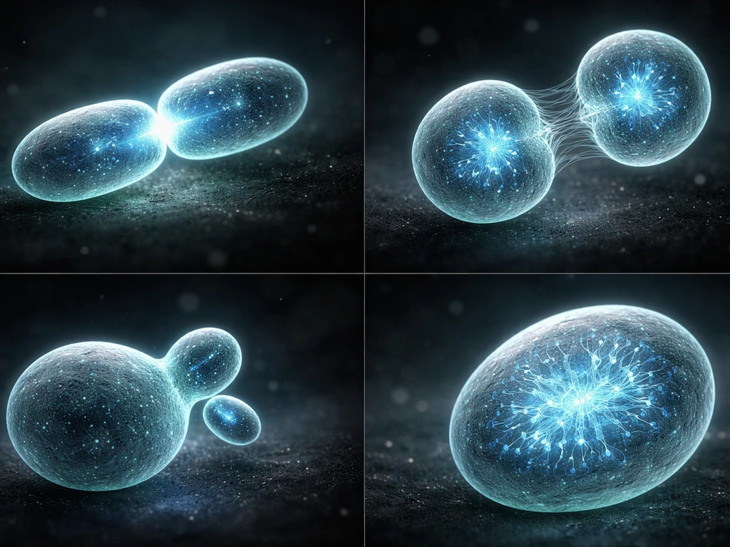
The cell-cycle control system: phases, engines, and checkpoints
The cell cycle runs through four phases: G1 (first gap, where the cell grows and preps), S phase (DNA replication), G2 (second gap, growth and repair continue), and M phase (mitosis, the actual division). The engine driving progression through these phases is a set of proteins called cyclin-dependent kinases, or Cdks, paired with their cyclin partners. Cyclins are the regulatory subunits that switch the Cdks on, and critically, different cyclin-Cdk combinations are active at different points in the cycle.
Cyclin D paired with Cdk4 or Cdk6 is the key driver in early G1. Cyclin E and Cdk2 push the cell through the G1-to-S transition. Cyclin A-Cdk2 handles S phase, and Cyclin B paired with Cdc2 (also called Cdk1) triggers entry into mitosis. These complexes don't just accelerate the cell forward; they also phosphorylate specific target proteins that unlock the next phase's machinery.
The checkpoints act as built-in pause buttons at three main moments: the G1/S transition (often called the restriction point or Start), the G2/M transition, and the spindle assembly checkpoint inside mitosis itself. At each one, the cell asks the same basic question: is everything ready and safe to proceed? If the answer is no, checkpoint proteins suppress the relevant cyclin-Cdk activity and the cell waits, or in some cases permanently withdraws from the cycle into a resting state called G0.
Signals that tell cells to divide faster
From the outside, the most important accelerators are growth factors, which are signaling proteins secreted by neighboring cells, the immune system, or specialized glands. When a growth factor binds its receptor on the cell surface, it triggers a signaling cascade (often through the RAS-MAPK or PI3K-AKT pathways) that boosts expression of Cyclin D. More Cyclin D means more active Cyclin D-Cdk4/6 complexes, which phosphorylate a protein called retinoblastoma protein (pRb). Phosphorylated pRb releases a transcription factor called E2F, which then switches on dozens of genes needed for DNA replication. In short, growth factors are the green light.
If growth factors are withdrawn before the cell crosses the restriction point in G1, Cyclin D levels drop rapidly and pRb stays in its repressive, dephosphorylated state. The cell can't activate E2F, can't initiate DNA replication, and slides into G0 quiescence instead. This is a normal, reversible state. Many cells in your body, like quiescent stem cells or resting immune cells, spend most of their lives in G0 and only re-enter the cycle when signaled.
Internally, nutrients and energy are equally critical. The mTOR (mechanistic target of rapamycin) pathway integrates signals about amino acid availability, glucose, and overall ATP levels. When nutrients are abundant, mTOR is active and drives protein synthesis and cell mass accumulation. When nutrients are scarce or ATP levels drop (triggering AMPK activation), mTOR is suppressed and growth slows or stops. Oxygen availability feeds into this too: low oxygen (hypoxia) activates HIF-1 transcription factors that broadly rewire cell metabolism and can inhibit proliferation.
The brakes: what stops or slows division
DNA damage is the most powerful brake the cell has. When sensors detect double-strand breaks or stalled replication forks, they activate kinases called ATM and ATR, which in turn stabilize and activate p53, one of the most important tumor suppressor proteins in biology. p53 then drives expression of a small protein called p21.
And p21 is a remarkable brake: it inhibits multiple cyclin-Cdk complexes at once, including Cyclin D-Cdk4/6 (the G1 engine), Cyclin E/A-Cdk2 (the S-phase engine), and Cyclin B1-Cdc2 (the mitotic entry complex). By hitting all three, p21 can enforce arrest at G1/S and prevent premature mitotic entry simultaneously.
NCBI Bookshelf notes that p21, a p53 target, inhibits multiple cyclin-Cdk complexes, including Cyclin D, Cdk4/6 and Cyclin E/A, Cdk2, by affecting E2F release from pRb repression, contributing to G1/S checkpoint arrest [p21, a p53 target, inhibits multiple cyclin-Cdk complexes including Cyclin D–Cdk4/6 and Cyclin E/A–Cdk2](https://www. ncbi. nlm. nih.
gov/books/NBK6412/). This gives the cell time to repair damage before replicating or dividing.
Replication stress, which happens when DNA polymerase stalls or replication forks collapse, activates the ATR-CHK1 signaling axis specifically. CHK1 phosphorylates and inactivates the CDC25 phosphatases that normally activate the cyclin-Cdk complexes needed for S-phase progression and G2/M transition. The result is a stalled S phase or a G2 arrest until the stress is resolved.
Broader cellular stress responses also act as brakes. Oxidative stress, heat shock, osmotic stress, and endoplasmic reticulum stress all feed into stress-activated kinase pathways that can suppress cyclin expression, promote Cdk inhibitor activity, or even activate the intrinsic apoptosis pathway if damage is irreparable. The cell is constantly weighing: fix it, pause it, or eliminate it.
Physical constraints: size, space, and the speed limit on growth
Even with all the right signals present, physics sets a hard ceiling on how fast a cell can grow and divide. The fundamental problem is the surface-area-to-volume ratio. As a spherical cell doubles in diameter, its volume increases by a factor of eight, but its surface area only increases by a factor of four. The plasma membrane is the cell's import/export interface, and if the ratio gets too unfavorable, the cell simply can't move nutrients in and waste products out fast enough to sustain the interior. This is why cells divide rather than just keep growing indefinitely.
There's also a DNA-to-cytoplasm ratio that cells monitor. Yeast cells have well-characterized size checkpoints: they won't commit to Start (the equivalent of the G1/S transition) unless they've accumulated enough mass relative to their DNA content. Similar mechanisms appear in mammalian cells, though the molecular details are still being worked out. The core idea is that the cell has molecular sensors that gauge whether it's big enough to divide.
Spatial crowding adds another constraint in tissues. Epithelial cells, for example, receive mechanical signals through integrin receptors and cell-adhesion molecules when they're packed tightly against neighbors. This contact inhibition suppresses proliferative signaling. Mechanosensitive pathways, including the Hippo pathway (which regulates the YAP/TAZ transcriptional co-activators), detect crowding and downregulate growth-promoting gene expression. So a cell's physical environment literally talks to its division machinery.
When control fails: cancer as a system breakdown
Cancer is what happens when the layered control system described above gets dismantled piece by piece through mutations. The most common mutations in cancer hit the accelerators or the brakes directly. RAS mutations (found in about 30% of all human cancers) lock the growth-factor signaling pathway in the permanently ON position, so Cyclin D stays elevated even without growth factors. Loss-of-function mutations in the retinoblastoma gene (pRb) remove the restriction point gatekeeper entirely, letting E2F run unchecked. Mutations in p53 (present in over 50% of human cancers) cripple the p21-mediated damage checkpoint, so cells with broken DNA keep dividing instead of arresting or dying.
What makes cancer particularly dangerous from a systems perspective is that it's usually not one broken layer but several. A typical cancer cell has bypassed external signal requirements (growth factor independence), silenced internal checkpoints (p53 loss, pRb loss), overcome size and contact inhibition signals (Hippo pathway mutations, integrin signaling changes), and sometimes even activated telomerase to bypass the replicative limit imposed by telomere shortening. Each broken layer on its own might not be enough. But stack them, and you get unchecked proliferation.
Understanding this layered logic is also why cancer therapy is so difficult. Targeting one broken component often isn't enough if the cell has already lost several backup brakes. And it's why the field is moving toward combination therapies that hit multiple control points simultaneously.
How to figure out what's actually limiting division in a real scenario
If you're a student, educator, or researcher trying to apply this knowledge to a specific situation, the most useful approach is to work through the three control layers systematically and ask which one is most likely the bottleneck.
| Control layer | What to look for | Key markers or measurements |
|---|---|---|
| External signals (growth factors, nutrients, oxygen) | Are the right signals present? Is the medium serum-depleted? Is oxygen or glucose low? | Cyclin D levels, pRb phosphorylation status, mTOR activity (phospho-S6K), AMPK activation |
| Internal checkpoints (DNA damage, replication stress) | Is DNA damaged or replication stalled? | γH2AX foci (DNA damage), p53 and p21 protein levels, CHK1/CHK2 phosphorylation, S-phase index by BrdU/EdU labeling |
| Physical/mechanical constraints (size, crowding) | Is the culture at high density? Is cell size unusually small or large? | Cell size by flow cytometry (forward scatter), YAP/TAZ nuclear localization, confluence of the culture, contact-inhibition assays |
In practice, start by asking whether cells are cycling at all. Flow cytometry with propidium iodide staining gives you a cell-cycle distribution in under an hour: you'll see peaks at G1 (2N DNA content), G2/M (4N), and a spread in between for S phase. If cells are piled up in G1, the restriction point is likely blocked, and you'd check Cyclin D levels and pRb phosphorylation. If they're arrested in G2/M, look for DNA damage markers or spindle checkpoint activation. If S phase is prolonged and the BrdU incorporation rate is low, replication stress is a likely culprit.
For a study or homework scenario, map the given conditions to this framework the same way. A question that says cells were grown in serum-free medium and stopped dividing is pointing directly at growth-factor withdrawal and Cyclin D loss. A question about cells treated with UV radiation points at the p53-p21-checkpoint axis. A question about cells in a densely packed tissue layer is pointing at contact inhibition and Hippo pathway signaling. Once you can identify the layer, you can reason through the downstream consequences.
A practical checklist for mapping any scenario
- Identify the starting condition: what changed in the cell's environment or what mutation is present?
- Determine which layer is affected: external signal, internal checkpoint, or physical constraint?
- Trace the molecular path: which cyclin-Cdk complex is affected, and at which phase does the block occur?
- Predict the observable outcome: G1 arrest, G2 arrest, slowed S phase, or exit into G0?
- If designing an experiment, choose a marker that directly reads out the relevant layer (see table above).
It's also worth knowing that these layers interact. A cell stressed by low nutrients (external layer) may activate AMPK, which can phosphorylate p53 independently of DNA damage, engaging the checkpoint layer even without any broken DNA. Understanding how the layers talk to each other is what separates a surface-level understanding from genuinely being able to predict what a cell will do.
Related questions worth exploring alongside this one include how specific gene types (proto-oncogenes and tumor suppressors) directly regulate cycle entry, whether cells always grow in size before committing to division, why some cell types opt to divide rather than grow bigger, and whether all cell types use the same checkpoint logic or have specialized versions of these controls. Each of those threads connects back to the same core framework covered here, just from a different angle.
FAQ
What is the practical difference between cell cycle arrest and entering quiescence (G0)?
Arrest usually means the cell remains capable of resuming the cycle once the block is removed (for example, temporary checkpoint activation). Quiescence (G0) is typically a more sustained resting program, where many cells reduce cyclin-Cdk activity broadly and depend on external signals and nutrients to re-enter. The key practical cue is reversibility and which checkpoint program is engaged.
How do researchers tell whether a block is at G1/S, G2/M, or during mitosis in experiments?
Beyond measuring DNA content, you can combine markers. For G1/S, look at cyclin D/pRb phosphorylation and whether E2F targets are active. For G2/M, check DNA damage and replication stress markers (for example, ATR-CHK1 pathway activity) and changes in CDC25 function. For mitosis-specific delay, spindle checkpoint readouts and mitotic spindle markers help distinguish a spindle arrest from a DNA damage arrest.
If growth factors are present, why might a cell still stop dividing?
Growth factors are a green light, but they cannot override internal brakes. Common reasons include DNA damage activating p53-p21, replication stress engaging ATR-CHK1, low ATP or amino acid scarcity suppressing mTOR, hypoxia activating HIF-1, or crowding/contact inhibition engaging mechanosensitive growth downregulation. In practice, identify the dominant constraint, not just the presence or absence of growth factor.
Can a cell divide without going through a normal S phase or mitosis sequence?
Generally, productive division requires completed DNA replication and successful mitotic segregation. If replication is incomplete or checkpoints are bypassed, cells can attempt aberrant mitosis, leading to genomic instability and often cell death or senescence. The safety system is largely checkpoint-dependent, so skipping steps usually increases missegregation risk rather than producing normal daughter cells.
Why do some cells appear to grow quickly or divide faster than others even with similar signals?
Different cell types have different baseline cyclin-Cdk levels, different sensitivities to growth-factor signaling, and distinct checkpoint thresholds. Also, their physical and metabolic context differs, such as nutrient access, oxygen levels, and how crowded the tissue environment is. Faster cycling can reflect both molecular wiring and microenvironmental constraints.
What happens if the cell reaches the “size needed” but nutrients are suddenly withdrawn?
Size accumulation depends on nutrients via pathways like mTOR, and cyclin abundance depends on growth-factor-driven transcriptional programs. If nutrients drop before the cell fully commits past restriction, mTOR suppression and energy stress (often via AMPK) can prevent the completion of the drive toward DNA replication. The likely outcome is a reversible halt or exit to a resting state rather than immediate division.
Does contact inhibition only apply to epithelial cells, or can other cell types experience similar effects?
While epithelial contact inhibition is well-studied (including integrin- and Hippo/YAP-TAZ-related effects), other adherent cell types also respond to mechanical cues and crowding. The exact molecular players can vary, but the decision logic often includes sensing of tissue density, adhesion, and mechanical stress that feeds into growth-promoting transcription.
How does replication stress differ from general DNA damage in terms of which checkpoint dominates?
Replication stress specifically involves stalled replication forks or collapsed forks, which strongly engages ATR-CHK1 signaling and suppresses CDC25 activity to delay S-phase completion and G2/M transition. General double-strand break damage more directly engages ATM and can strongly route through p53-p21. Both can produce arrest, but the dominant pathway and timing can differ.
Is losing p53 always enough to cause uncontrolled division, or can cells still be restrained?
Loss of p53 removes a major brake, but other restraints often remain, such as pRb control at G1/S, checkpoint signaling from replication stress, and metabolic or contact-inhibition limits. Uncontrolled proliferation in cancer usually involves multiple layers failing simultaneously, which is why targeting one component can be insufficient.
Why do cancer cells often require multiple mutations instead of just one?
A single mutation may accelerate one step, but other brakes can still halt the cycle. For example, growth-factor independence needs checkpoint bypass to tolerate damaged DNA, and bypassing pRb restriction often must coincide with disabling p53-p21 for replication stress to be tolerated. Physical and telomere-related limits also add constraints, so stacked defects commonly produce robust proliferation.
In cell-cycle profiling, what common mistake leads to misinterpreting “more in G1” as “slower division”?
A higher fraction in G1 can reflect true G1/S arrest, but it can also come from changes in cell death rate, altered DNA staining assumptions, or selective survival of certain subpopulations. Complement DNA content with viability controls and appropriate marker staining (and consider gating strategy) to avoid mistaking survival or technical effects for a specific checkpoint block.
Next Article
Do All Cells Grow and Divide in the Same Way?
Cells grow similarly but divide differently by type and organism, including mitosis, meiosis, binary fission, and endore